Vestibulo Cochlear Nerve and Your Heart

As someone who has personally experienced the impact of vestibulo cochlear nerve disorders on cardiovascular health, I understand the importance of shedding light on this often overlooked connection. In this article, we will delve into the intricate relationship between the vestibulo cochlear nerve and your heart, exploring its anatomy, functions, symptoms, diagnosis, treatment options, and exciting advancements in research.

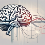
Understanding the Vestibulo Cochlear Nerve

The vestibulo cochlear nerve, also known as the eighth cranial nerve, comprises two distinct components: the vestibular nerve and the cochlear nerve. Working in harmony, these nerves play crucial roles in our sense of balance, spatial orientation, and auditory perception.

Anatomy of the Vestibulo Cochlear Nerve

The vestibulo cochlear nerve originates deep within the inner ear and travels along the temporal bone, connecting the delicate structures responsible for balance, such as the vestibular apparatus, with the auditory pathways of the brain. This intricate web of nerve fibers ensures seamless communication between our balance system and hearing capabilities, enabling us to navigate the world around us.

Furthermore, the vestibulo cochlear nerve is intricately connected to the vestibular system, which includes the semicircular canals and otolithic organs. These structures are essential for detecting changes in head position and movement, providing vital information to the brain about our spatial orientation. The vestibular nerve, in particular, plays a key role in maintaining our equilibrium and coordinating eye movements to compensate for head motion.

Function of the Vestibulo Cochlear Nerve

Now, let’s dive deeper into the fascinating functions of the vestibulo cochlear nerve. Firstly, the vestibular nerve provides crucial information to the brain about the position and movement of our head in space. This information allows for the coordination of our muscles and helps us maintain balance, whether we are standing still or engaged in dynamic activities. Secondly, the cochlear nerve facilitates the transmission of sound signals from the inner ear to the brain, enabling us to perceive and interpret a rich tapestry of auditory stimuli.

Moreover, the vestibulo cochlear nerve is responsible for encoding the intensity and frequency of sound waves, allowing us to differentiate between various pitches and volumes. This intricate process involves the conversion of mechanical vibrations in the cochlea into electrical signals that can be processed by the brain. The precise coordination between the vestibular and cochlear components of the nerve ensures that we can not only hear but also maintain our balance in response to auditory cues.

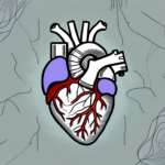
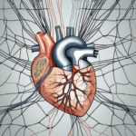
The Connection between the Vestibulo Cochlear Nerve and the Heart

Surprisingly, recent research has shed light on the profound relationship between the vestibulo cochlear nerve and our cardiovascular system. Studies have revealed that this delicate nerve network not only influences our balance and hearing but also plays a role in the regulation of heart function.

Understanding the intricate connection between the vestibulo cochlear nerve and the heart opens up a fascinating realm of possibilities in both neurological and cardiological research. The nerve, responsible for transmitting crucial sensory information from the inner ear to the brain, is now recognized for its additional role in maintaining cardiovascular homeostasis.

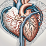
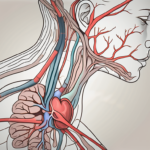
Role of the Vestibulo Cochlear Nerve in Cardiovascular Function

The vestibulo cochlear nerve, through its intricate connections and interactions with the brainstem and autonomic nervous system, contributes to the regulation of heart rate, blood pressure, and vascular tone. This means that any disruption or dysfunction within the nerve can potentially impact cardiovascular health, leading to a range of symptoms and complications.

Moreover, the vestibulo cochlear nerve’s involvement in cardiovascular function highlights the complexity of the body’s interconnected systems. The nerve’s ability to modulate heart rate and blood pressure showcases the intricate dance between sensory input and physiological response, emphasizing the holistic nature of human health.

The Impact of Vestibulo Cochlear Nerve Disorders on the Heart

Individuals experiencing vestibulo cochlear nerve disorders may unknowingly face heightened risks to their heart health. Conditions such as vestibular migraine, Meniere’s disease, and vestibular schwannoma can disturb the delicate balance within this nerve network, triggering symptoms such as dizziness, vertigo, and hearing loss. Moreover, these disorders can also indirectly affect the heart, leading to fluctuations in blood pressure, increased cardiovascular strain, and even potential risk factors for cardiovascular diseases.

By delving deeper into the relationship between the vestibulo cochlear nerve and the heart, researchers aim to not only enhance our understanding of these interconnected systems but also pave the way for novel treatment approaches that target both neurological and cardiovascular aspects. The intricate interplay between the nerve and the heart serves as a reminder of the body’s remarkable complexity and the ongoing quest to unravel its mysteries.

Symptoms and Diagnosis of Vestibulo Cochlear Nerve Disorders

Recognizing the symptoms and obtaining an accurate diagnosis are vital steps towards effectively managing vestibulo cochlear nerve disorders and safeguarding your heart health.

Vestibulo cochlear nerve disorders, also known as cranial nerve VIII disorders, can have a significant impact on an individual’s quality of life. These disorders affect the vestibular and auditory systems, leading to a range of debilitating symptoms that can disrupt daily activities and overall well-being.

Common Symptoms of Vestibulo Cochlear Nerve Disorders

The symptoms of vestibulo cochlear nerve disorders can manifest differently in each individual, but some common signs include intense dizziness, recurrent vertigo episodes, imbalance, tinnitus (ringing in the ears), and hearing loss. It is important to seek medical attention if you experience any of these concerning symptoms.

Dizziness and vertigo, in particular, can be distressing and may significantly impact an individual’s ability to perform tasks that require balance and coordination. The sensation of spinning or feeling off-balance can be triggered by various factors, including changes in head position, sudden movements, or even just getting out of bed in the morning.

Diagnostic Procedures for Vestibulo Cochlear Nerve Disorders

Diagnosing vestibulo cochlear nerve disorders typically involves a comprehensive evaluation of your medical history, a physical examination, and various specialized tests. These tests can include audiometry, electronystagmography, vestibular evoked myogenic potential testing, and imaging studies like MRI or CT scans. Consulting with an experienced medical professional is essential to ensure a proper diagnosis.

During the diagnostic process, healthcare providers may also assess the impact of vestibulo cochlear nerve disorders on an individual’s emotional well-being and cognitive function. The interconnected nature of the vestibular and auditory systems means that these disorders can have far-reaching effects beyond just physical symptoms, potentially leading to anxiety, depression, and difficulties with concentration and memory.

Treatment and Management of Vestibulo Cochlear Nerve Disorders

While there is no one-size-fits-all approach to treating vestibulo cochlear nerve disorders, there are several medical and lifestyle interventions that can help alleviate symptoms and improve overall quality of life.

Understanding the complexities of vestibulo cochlear nerve disorders is essential in developing an effective treatment plan. These disorders can stem from various underlying causes, such as Meniere’s disease, vestibular neuritis, acoustic neuroma, or even certain viral infections. Each condition may require a tailored approach to address the specific symptoms and challenges faced by the individual.

Medical Treatments for Vestibulo Cochlear Nerve Disorders

Medications, such as anti-vertigo medications, diuretics, and migraine preventatives, are commonly prescribed to manage symptoms like dizziness, vertigo attacks, and associated conditions. Additionally, vestibular rehabilitation therapy, a specialized form of physical therapy, can also provide significant benefits by helping to retrain the brain to adapt and promote balance.

In some cases, surgical interventions may be necessary to address structural issues affecting the vestibulo cochlear nerve. Procedures like vestibular nerve section or decompression surgery can help alleviate pressure on the nerve and improve symptoms in certain individuals. These interventions are typically considered when conservative treatments have not provided sufficient relief.

Lifestyle Changes to Manage Vestibulo Cochlear Nerve Disorders

Beyond medical treatments, adopting certain lifestyle modifications can play a crucial role in managing vestibulo cochlear nerve disorders and promoting heart health. These lifestyle changes can include stress reduction techniques, a balanced diet low in sodium, regular exercise, proper hydration, and adequate rest. However, it is crucial to consult with healthcare professionals to tailor these changes to your specific condition and needs.

Furthermore, incorporating mindfulness practices, such as meditation or yoga, can help individuals cope with the emotional and psychological impact of living with vestibulo cochlear nerve disorders. These practices promote relaxation, reduce anxiety levels, and improve overall mental well-being, which are all essential components of a holistic treatment approach.

The Future of Vestibulo Cochlear Nerve Research

Exciting advancements in vestibulo cochlear nerve research offer hope for improved understanding, management, and potential breakthroughs in the future.

The field of vestibulo cochlear nerve research is constantly evolving, with scientists and researchers pushing the boundaries of knowledge to unlock new possibilities. One area of focus is the development of innovative treatment approaches, such as gene therapy and regenerative medicine. These cutting-edge initiatives aim to target and repair damaged vestibulo cochlear nerve pathways, paving the way for potential restoration of balance and reduction of symptoms. Additionally, these advancements hold promise for preserving cardiovascular health, as the vestibulo cochlear nerve has been found to play a role in maintaining optimal heart function.

Advances in Vestibulo Cochlear Nerve Treatment

Scientists and researchers continue to explore innovative treatment approaches, such as gene therapy and regenerative medicine, to target and repair damaged vestibulo cochlear nerve pathways. These groundbreaking initiatives hold promise for restoring balance, reducing symptoms, and preserving cardiovascular health.

Gene therapy, for example, involves the introduction of specific genes into the body to correct genetic abnormalities that may be contributing to vestibulo cochlear nerve disorders. This approach has shown great potential in preclinical studies, with researchers successfully restoring balance and reducing symptoms in animal models. Regenerative medicine, on the other hand, focuses on harnessing the body’s own regenerative capabilities to repair damaged nerve pathways. By utilizing stem cells or other regenerative techniques, scientists are working towards developing therapies that can promote the growth and repair of vestibulo cochlear nerve cells.

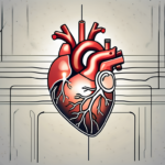
Potential Implications for Heart Health

Further research into the mechanisms underlying the intricate connection between the vestibulo cochlear nerve and the heart may reveal exciting opportunities for early detection, prevention, and targeted interventions for cardiovascular diseases. By unraveling the mysteries of this interplay, we might uncover new approaches to improve heart health and enhance overall well-being.

Recent studies have shown that the vestibulo cochlear nerve is not only involved in maintaining balance and hearing, but also plays a role in regulating cardiovascular function. The nerve fibers that make up the vestibulo cochlear nerve have been found to have direct connections to the heart, influencing its rhythm and overall performance. This intriguing link has sparked interest among researchers, who are now investigating whether abnormalities in the vestibulo cochlear nerve could be early indicators of cardiovascular diseases. By identifying these potential biomarkers, medical professionals may be able to detect and intervene in heart conditions at an earlier stage, leading to more effective treatments and improved outcomes.

Furthermore, understanding the intricate relationship between the vestibulo cochlear nerve and the heart could open up new avenues for targeted interventions. By developing therapies that specifically target the nerve pathways involved in cardiovascular regulation, researchers may be able to design treatments that not only address vestibulo cochlear nerve disorders but also have a positive impact on heart health. This integrated approach has the potential to revolutionize the field of cardiovascular medicine, offering new hope for individuals at risk of or already living with heart conditions.

The Brain Stimulator and Your Heart

I never realized the connection between the brain and the heart until I stumbled upon an intriguing topic during my research. This led me down a fascinating rabbit hole, uncovering the impact of brain stimulation on heart health. It’s incredible how our bodies work in unison, with one organ affecting another more than we could have ever imagined. So, let’s dive into the world of brain stimulation and its relationship with your heart.

Understanding the Connection Between the Brain and Heart

Our bodies are complex machines, intricately interconnected and constantly communicating with one another. In the case of the brain and the heart, this connection is particularly profound. It all begins with the autonomic nervous system, a regulatory network that controls our involuntary bodily functions, including heart rate and blood pressure.

The Role of the Autonomic Nervous System

The autonomic nervous system consists of two branches: the sympathetic and parasympathetic systems. The sympathetic system acts as a gas pedal, increasing heart rate and blood pressure, while the parasympathetic system acts as a brake, slowing down the heart rate and promoting relaxation.

These two branches of the autonomic nervous system work in harmony, striving to maintain a delicate balance. However, factors like stress, emotional upheaval, or certain medical conditions can disrupt this equilibrium and impact heart health.

The Impact of Emotions on Heart Health

We’ve all experienced it – the rapid heartbeat when we’re excited, the heavy feeling on our chest when we’re anxious. These emotional responses are not just fleeting sensations; they can have a profound impact on our heart health.

Research has shown that chronic stress and negative emotions, if left unchecked, can increase the risk of heart disease. The brain, with its intricate web of emotions, plays a central role in this connection. When we’re stressed or experiencing intense emotions, the brain signals the release of stress hormones, such as adrenaline and cortisol. These hormones, in turn, can increase heart rate, elevate blood pressure, and strain our cardiovascular system.

But what about positive emotions? Can they have a beneficial effect on our heart health? The answer is a resounding yes! Studies have shown that positive emotions, such as joy, love, and gratitude, can have a protective effect on the heart. When we experience these positive emotions, the brain releases endorphins and other feel-good chemicals that can lower heart rate, reduce blood pressure, and improve overall cardiovascular health.

Furthermore, the brain-heart connection goes beyond emotions. The brain also plays a crucial role in regulating our sleep patterns, which in turn can impact heart health. Lack of sleep or poor sleep quality can lead to an increased risk of heart disease, as it disrupts the body’s natural rhythm and puts additional strain on the cardiovascular system.

So, it’s clear that the connection between the brain and heart is not just a simple one. It’s a complex interplay of emotions, hormones, and sleep patterns. By understanding this connection and taking steps to manage stress, cultivate positive emotions, and prioritize healthy sleep habits, we can support the well-being of both our brain and heart.

The Science Behind Brain Stimulators

Now that we have a grasp of the intertwined nature of the brain and heart, let’s delve into the science behind brain stimulators and how they could potentially aid in maintaining heart health.

Understanding the intricate relationship between the brain and the heart is crucial in exploring the potential benefits of brain stimulators in cardiovascular health. The brain plays a significant role in regulating heart function through the autonomic nervous system, which controls involuntary actions like heart rate and blood pressure. By targeting specific brain regions with stimulation, researchers aim to modulate these autonomic functions and potentially improve overall heart health.

The Process of Brain Stimulation

Brain stimulation involves the use of targeted electrical pulses or magnetic fields to modulate specific areas of the brain. It’s a non-invasive procedure that aims to regulate neuronal activity and improve brain function. This technique has been widely used in the treatment of various neurological and psychiatric conditions, such as Parkinson’s disease and depression.

When brain stimulators are applied, they can trigger changes in neurotransmitter levels, synaptic connections, and neural plasticity, leading to alterations in brain activity. By harnessing these mechanisms, researchers hope to not only address neurological disorders but also explore the potential impact on cardiovascular function.

Different Types of Brain Stimulators

There are different types of brain stimulators, each with its unique methods and applications. Transcranial magnetic stimulation (TMS), for instance, uses magnetic fields to stimulate specific brain regions from outside the body. Another technique, transcranial direct current stimulation (tDCS), involves the application of weak electrical currents to modulate brain activity.

These brain stimulation techniques hold promise not just in the realm of mental health but also in the vast landscape of heart health. By influencing certain areas of the brain, researchers believe that it’s possible to regulate heart rhythm and potentially lower the impact of stress on the cardiovascular system.

As research in this field progresses, the potential for innovative therapies that combine brain stimulation and cardiovascular health management continues to expand. By unraveling the complex interplay between the brain, heart, and overall physiological well-being, scientists aim to pave the way for novel approaches in enhancing both mental and cardiovascular resilience.

Potential Benefits of Brain Stimulation for Heart Health

The notion of using brain stimulation to benefit heart health might sound like something out of a science fiction movie. However, ongoing research indicates that it could be a viable strategy for improving cardiovascular well-being.

Regulating Heart Rhythm Through Brain Stimulation

One of the key areas of interest in brain stimulation research is its potential to regulate heart rhythm. A healthy heart relies on a synchronized rhythm to pump blood efficiently throughout the body. Studies have shown that specific brain regions, when stimulated, can influence the autonomic nervous system and restore heart rhythm to a more desirable state.

For instance, researchers have focused on the role of the hypothalamus, a region deep within the brain, in controlling heart rate. By using targeted brain stimulation techniques, scientists have been able to modulate the activity of the hypothalamus, leading to improved heart rhythm regulation. This breakthrough has opened up new possibilities for managing heart rhythm disorders and enhancing overall cardiovascular health.

Stress Reduction and Heart Health

Another significant benefit of brain stimulation is its potential to reduce stress levels, which can have a direct impact on heart health. Chronic stress takes a toll on our bodies, including our cardiovascular system. By targeting specific brain areas involved in stress regulation, brain stimulators may minimize the negative impact of stress on the heart.

Imagine a world where stressors no longer seize control of our physiological well-being. While it may sound like a distant dream, advancements in brain stimulation research are bringing us one step closer to this reality.

Moreover, recent studies have shown that brain stimulation techniques, such as transcranial magnetic stimulation (TMS), can activate the release of endorphins and other feel-good neurotransmitters in the brain. These natural chemicals not only promote a sense of well-being but also have a positive impact on heart health. By stimulating the brain’s reward centers, brain stimulation may help counteract the negative effects of stress and improve overall cardiovascular function.

Furthermore, researchers are exploring the potential of brain stimulation in promoting the growth of new blood vessels in the heart, a process known as angiogenesis. By stimulating specific brain regions, scientists have observed an increase in the release of growth factors that can stimulate the formation of new blood vessels. This exciting finding could have significant implications for individuals with heart disease or those recovering from a heart attack, as it could potentially enhance blood flow to damaged areas and aid in the healing process.

Risks and Considerations of Brain Stimulation

Like any medical intervention, brain stimulation also comes with its fair share of risks and considerations that need to be carefully weighed.

When delving into the realm of brain stimulation, it’s essential to understand the intricate balance between potential benefits and possible risks. While the field of neuroscience has made significant advancements in this area, there are still nuances to be explored to ensure the utmost safety and efficacy of these procedures.

Potential Side Effects of Brain Stimulation

Although brain stimulation techniques are generally considered safe, there can be side effects. These can range from mild headaches or scalp discomfort during the procedure to more severe effects such as seizures or mood changes.

Furthermore, the impact of brain stimulation on long-term cognitive function and emotional well-being is an area of ongoing research and debate. It is crucial for both healthcare providers and patients to remain vigilant in monitoring and reporting any unexpected or concerning side effects that may arise post-treatment.

It’s important to note that these adverse reactions are relatively rare and are typically closely monitored by healthcare professionals. Nevertheless, it’s crucial to consult with a qualified medical specialist before considering brain stimulation as a potential avenue for improving heart health.

Who Should Avoid Brain Stimulation?

While brain stimulation holds promise, it might not be suitable for everyone. Individuals with certain medical conditions, such as epilepsy or a history of seizures, may not be ideal candidates for this procedure. Additionally, pregnant women or those with implanted medical devices, such as pacemakers, should exercise caution and seek expert medical advice before pursuing brain stimulation as a potential therapy.

Moreover, the personalized nature of brain stimulation treatments underscores the importance of a thorough evaluation by a multidisciplinary team of healthcare professionals. Factors such as individual medical history, current medications, and lifestyle choices can all play a role in determining the suitability and potential risks associated with brain stimulation.

Future Research on Brain Stimulation and Heart Health

The field of brain stimulation is still in its infancy, with much more to be explored and understood. As with any emerging scientific domain, there are current limitations and exciting potential developments worth considering.

Current Limitations of Research

While preliminary studies have shown promising results, the current body of research on brain stimulation and heart health is relatively limited. Further well-designed clinical trials are necessary to confirm the efficacy and safety of these techniques, as well as to identify the optimal parameters and protocols for the desired outcomes.

One particular limitation is the need for long-term follow-up studies. Many of the existing research focuses on short-term effects, but it is crucial to understand the potential long-term benefits and risks associated with brain stimulation for heart health. This information will be vital in determining the sustainability and durability of these interventions.

Another challenge researchers face is the customization of brain stimulation treatments. Everyone’s brain is unique, and what works for one person may not necessarily work for another. Tailoring these therapies to individual needs is an area that requires extensive investigation and fine-tuning. Factors such as age, gender, and underlying health conditions may influence the response to brain stimulation, and understanding these nuances will be crucial in optimizing treatment outcomes.

Potential Developments in the Field

Despite the challenges, the field of brain stimulation and heart health is rife with potential. Advances in technology and a deeper understanding of brain-heart interactions could pave the way for targeted therapies that are more precise, effective, and accessible to a broader population.

Furthermore, ongoing research exploring the combination of brain stimulation with other modalities, such as pharmaceutical interventions or lifestyle modifications, holds tremendous promise in optimizing heart health outcomes. By combining the power of brain stimulation with other evidence-based approaches, researchers may be able to unlock synergistic effects that could lead to even greater improvements in cardiovascular health.

Moreover, investigating the potential of brain stimulation in different populations, such as individuals with specific heart conditions or those at different stages of heart disease, could provide valuable insights into the effectiveness of these interventions across diverse patient groups. This knowledge could help tailor treatment approaches and improve outcomes for individuals with varying heart health needs.

Conclusion

When it comes to our health, it’s essential to view our bodies holistically. The brain and the heart, two of our most vital organs, share a deep connection that extends far beyond what meets the eye. Brain stimulation, with its potential to influence heart health, offers a tantalizing avenue for future medical advancements.

While the journey towards harnessing the full potential of brain stimulation is still underway, the prospect of a world where brain and heart work in perfect harmony is becoming increasingly tangible. As research progresses, we may witness groundbreaking therapies that could revolutionize the way we approach heart health and enhance our overall well-being.

Vagus Nerve Stimulators and Your Heart

The vagus nerve is a remarkable part of our body that plays a vital role in regulating various functions, including our heart rate and rhythm. In recent years, there has been increasing interest in using vagus nerve stimulation as a therapeutic approach for improving heart health. In this article, we will explore the intricate connection between the vagus nerve and the heart, the potential benefits of vagus nerve stimulation, as well as the risks and side effects associated with this modality.

Understanding the Vagus Nerve

Before delving into the details of vagus nerve stimulation, it is essential to have a basic understanding of the vagus nerve itself. The vagus nerve, also known as the tenth cranial nerve, is one of the longest and most complex nerves in our body. It connects the brain to various organs, including the heart, lungs, and digestive system.

The vagus nerve plays a crucial role in the parasympathetic nervous system, which is responsible for the body’s rest and digest response. This nerve is a key player in regulating involuntary bodily functions, such as heart rate, digestion, and respiratory rhythm. It is often referred to as the “wandering nerve” due to its extensive distribution throughout the body.

Anatomy of the Vagus Nerve

The vagus nerve originates in the brainstem and branches out to form a network of fibers that innervate different parts of the body. It consists of both sensory and motor fibers, allowing it to transmit signals to and from the brain.

As the vagus nerve travels through the body, it sends and receives information from various organs, helping to maintain homeostasis and regulate bodily functions. The nerve has multiple branches that reach the heart, lungs, liver, spleen, and intestines, among other organs. This widespread connectivity underscores its importance in overall health and well-being.

Functions of the Vagus Nerve

The vagus nerve is responsible for regulating several vital bodily functions, such as breathing, digestion, and heart function. It acts as a communication highway between the brain and the organs it innervates, constantly relaying information and coordinating responses.

In addition to its role in basic physiological processes, the vagus nerve also plays a crucial part in the mind-body connection. It is involved in the regulation of mood, stress responses, and even social bonding. Some researchers have dubbed the vagus nerve as the “nerve of compassion” due to its influence on empathy and emotional regulation.

The Role of Vagus Nerve in Heart Function

The vagus nerve exerts a profound influence on heart rate and rhythm. It acts as a natural regulatory system, helping to maintain a balanced and optimal state of cardiovascular function.

The vagus nerve, also known as the 10th cranial nerve, is the longest and most complex of the cranial nerves. It originates in the brainstem and travels through the neck to the thorax and abdomen, where it branches out to various organs, including the heart. This extensive network allows the vagus nerve to communicate vital information between the brain and the heart, playing a crucial role in maintaining homeostasis.

Vagus Nerve and Heart Rate

One of the primary functions of the vagus nerve is to slow down the heart rate when needed. It does this by releasing a neurotransmitter called acetylcholine, which binds to specific receptors in the heart and lowers the heart rate. This mechanism is crucial in preventing heart rate irregularities and optimizing cardiac performance.

Furthermore, the vagus nerve acts as a key player in the parasympathetic nervous system, the branch of the autonomic nervous system responsible for rest and digest functions. By modulating heart rate, the vagus nerve helps the body conserve energy during periods of relaxation and recovery.

Vagus Nerve and Heart Rhythm

In addition to heart rate control, the vagus nerve also plays a role in regulating the electrical activity of the heart. It helps maintain a coordinated heart rhythm by influencing the sinus node, a natural pacemaker in the heart. Through its intricate neural connections, the vagus nerve helps ensure that the heart beats in a synchronized and efficient manner.

Moreover, the vagus nerve’s influence on heart rhythm extends beyond the sinus node to other cardiac structures, such as the atrioventricular node and the ventricles. By fine-tuning the timing and sequence of electrical impulses, the vagus nerve contributes to the overall stability and effectiveness of the heart’s pumping action, supporting optimal circulation throughout the body.

Vagus Nerve Stimulators: An Overview

In cases where the natural regulation of the vagus nerve is disrupted, such as in certain heart conditions, vagus nerve stimulation can be used as a therapeutic approach. Vagus nerve stimulators are medical devices that deliver targeted electrical impulses to the vagus nerve, aiming to restore and optimize its function.

Understanding the intricate network of the vagus nerve and its role in regulating various bodily functions is crucial in appreciating the significance of vagus nerve stimulators. The vagus nerve, also known as the 10th cranial nerve, is a key player in the parasympathetic nervous system, responsible for controlling involuntary bodily processes such as heart rate, digestion, and respiratory function. When this nerve’s function is compromised, it can lead to a range of health issues, making interventions like vagus nerve stimulation essential in managing certain conditions.

How Vagus Nerve Stimulators Work

Vagus nerve stimulators are typically implanted under the skin, with electrodes attached to the vagus nerve. These devices deliver electrical impulses at pre-defined intervals, mimicking the natural signals of the vagus nerve. By modulating the electrical activity of the vagus nerve, these stimulators can help regulate heart rate, rhythm, and other functions under certain circumstances.

Moreover, the precise mechanism of action of vagus nerve stimulators involves the activation of specific pathways within the brain that are linked to mood regulation. This additional benefit has expanded the application of vagus nerve stimulation beyond cardiac conditions to include the management of treatment-resistant depression and other mood disorders.

Types of Vagus Nerve Stimulators

There are different types of vagus nerve stimulators available, each with its own unique features and settings. Some devices provide continuous stimulation, while others can be activated as needed. The choice of the specific device depends on the individual’s medical condition and the recommendations of their healthcare provider.

Furthermore, advancements in technology have led to the development of miniaturized vagus nerve stimulators that offer increased precision and customization in therapy delivery. These modern devices are equipped with sophisticated algorithms that can adapt stimulation parameters based on real-time physiological data, ensuring optimal outcomes for patients with varying needs.

Benefits of Vagus Nerve Stimulation for Heart Health

Vagus nerve stimulation has shown promising results in improving heart health and managing certain cardiovascular conditions. However, it is essential to note that the use of vagus nerve stimulators is typically reserved for specific cases and should always be done under medical supervision.

Understanding the intricate connection between the vagus nerve and heart health is crucial in exploring the potential benefits of vagus nerve stimulation. The vagus nerve, a key component of the parasympathetic nervous system, plays a significant role in regulating heart function, including heart rate and blood pressure. By modulating the activity of the vagus nerve through stimulation, it is believed that certain cardiovascular conditions can be positively influenced.

Vagus Nerve Stimulation and Heart Disease

Vagus nerve stimulation has been studied as a potential therapy for various cardiovascular conditions, including certain types of heart disease. Research suggests that it may have beneficial effects on heart function, such as reducing inflammation and improving overall cardiac performance. However, further studies are needed to fully understand its efficacy and safety in specific cardiac conditions.

One of the key mechanisms through which vagus nerve stimulation may benefit individuals with heart disease is by reducing the production of pro-inflammatory cytokines. These molecules play a role in promoting inflammation within the cardiovascular system, which can contribute to the progression of heart disease. By dampening this inflammatory response through vagus nerve stimulation, there is potential for mitigating the detrimental effects of chronic inflammation on the heart.

Vagus Nerve Stimulation and Heart Failure

Heart failure is a complex condition that requires comprehensive management. While vagus nerve stimulation has shown promise in some cases, it is not a standalone treatment for heart failure. It should be considered as a complementary therapy alongside other established interventions, such as medication, lifestyle modifications, and device implantation.

In individuals with heart failure, vagus nerve stimulation may offer benefits by helping to regulate autonomic balance. Imbalances in the autonomic nervous system, which controls involuntary functions such as heart rate and blood vessel constriction, are common in heart failure. By modulating the activity of the vagus nerve, which is a key regulator of the parasympathetic branch of the autonomic nervous system, it is possible to restore a more optimal balance between the sympathetic and parasympathetic systems, potentially improving heart function.

Risks and Side Effects of Vagus Nerve Stimulation

Although vagus nerve stimulation can have potential benefits, it is crucial to be aware of the associated risks and side effects. Understanding the intricacies of this procedure can help individuals make informed decisions about their health and well-being.

Vagus nerve stimulation is a therapeutic approach that involves the use of electrical impulses to regulate brain activity and treat various medical conditions. By targeting the vagus nerve, which plays a crucial role in the body’s autonomic nervous system, this treatment can offer relief for individuals struggling with epilepsy, depression, and other neurological disorders.

Common Side Effects

Common side effects of vagus nerve stimulation may include hoarseness, throat pain, coughing, shortness of breath, and neck pain. These side effects are usually temporary and tend to improve over time as the body adjusts to the stimulation. It is important for patients to communicate openly with their healthcare providers about any discomfort they may experience during the course of treatment.

In addition to the physical side effects, some individuals may also encounter emotional or psychological changes as a result of vagus nerve stimulation. These can range from mood swings and anxiety to improvements in overall well-being and cognitive function. Monitoring these changes and seeking support when needed is essential for a holistic approach to treatment.

Potential Risks and Complications

Like any medical procedure, vagus nerve stimulation carries some risks. These may include infection, nerve damage, difficulty swallowing, and vocal cord paralysis. It is essential to discuss the potential risks and benefits with a healthcare professional before considering vagus nerve stimulation as a treatment option. By weighing the pros and cons of this intervention, individuals can make educated decisions about their healthcare journey.

Furthermore, ongoing research is being conducted to explore the long-term effects of vagus nerve stimulation on various health conditions. By participating in clinical trials and staying informed about the latest advancements in medical technology, patients can contribute to the collective knowledge surrounding this innovative treatment approach.

Preparing for Vagus Nerve Stimulation Therapy

If vagus nerve stimulation is recommended as a therapeutic option, it is essential to be well-prepared and informed about what to expect. Understanding the intricacies of this therapy can help individuals feel more at ease and empowered throughout the process.

Vagus nerve stimulation therapy involves the use of a device that is implanted in the body to deliver electrical impulses to the vagus nerve, which plays a crucial role in regulating various bodily functions. This therapy is often recommended for individuals with epilepsy, depression, or other neurological conditions that have not responded well to traditional treatments.

What to Expect During the Procedure

The vagus nerve stimulation procedure is typically performed under general anesthesia. A small incision is made to implant the device and attach the electrodes to the vagus nerve. The surgery itself is considered minimally invasive, and most individuals can expect a relatively short recovery period. The electrodes deliver controlled electrical impulses to the vagus nerve, helping to regulate abnormal brain activity and improve symptoms over time.

During the procedure, healthcare providers will monitor vital signs and ensure the patient’s comfort and safety. It is normal to experience some soreness or discomfort at the implantation site following the surgery, but this can usually be managed with pain medication and proper wound care.

Post-Procedure Care and Recovery

After the vagus nerve stimulation device is implanted, regular follow-up appointments will be necessary to ensure its proper functioning and make any needed adjustments. It is important to closely follow the post-procedure care instructions provided by the healthcare team and report any concerns or complications promptly. Engaging in open communication with healthcare providers can help address any issues that may arise and optimize the effectiveness of the therapy.

Individuals undergoing vagus nerve stimulation therapy may also be advised to keep a journal to track their symptoms and any changes they notice after the device is implanted. This information can be valuable for healthcare providers in fine-tuning the therapy settings to achieve the best possible outcomes. Additionally, adopting a healthy lifestyle that includes regular exercise, adequate sleep, and stress management techniques can complement the benefits of vagus nerve stimulation therapy and support overall well-being.

Future of Vagus Nerve Stimulation in Cardiology

As medical research and technology continue to advance, the future of vagus nerve stimulation in cardiology looks promising.

Ongoing Research and Developments

Scientists and clinicians are continuously conducting research to explore the full potential of vagus nerve stimulation in improving heart health. Ongoing studies aim to uncover its mechanisms of action, refine its applications, and identify the patients who may benefit the most from this therapy.

Recent studies have shown that vagus nerve stimulation can not only help regulate heart rate and blood pressure but also modulate inflammation and improve overall cardiac function. This multifaceted approach highlights the potential of vagus nerve stimulation to address various aspects of cardiovascular health beyond traditional treatments.

Potential Future Applications in Heart Health

The future of vagus nerve stimulation may involve the development of more advanced devices and innovative treatment approaches. With further advancements, vagus nerve stimulation may become a more tailored and effective therapeutic option for specific cardiac conditions, potentially improving the quality of life for individuals with heart disease.

Furthermore, ongoing research is exploring the potential of vagus nerve stimulation in addressing arrhythmias, heart failure, and even cardiac arrest. By fine-tuning the parameters of stimulation and enhancing device technology, researchers aim to expand the scope of vagus nerve stimulation in cardiology to encompass a wider range of cardiovascular disorders.

In conclusion, vagus nerve stimulators offer a unique and promising approach to improving heart health. While they can provide benefits in certain cardiac conditions, it is crucial to work closely with healthcare professionals to determine the suitability of this therapy. The vagus nerve and its intricate connection to heart function continue to be an area of active research, paving the way for potential advancements in the field of cardiology.

Auricular Nerve and Your Heart

As someone who has experienced the fascinating connection between the auricular nerve and the heart, I am excited to share my knowledge with you. Understanding the interplay of these two vital components of our bodies can shed light on a range of health implications. So, let’s dive right in!

Understanding the Auricular Nerve

Before we explore the intricate relationship between the auricular nerve and the heart, it’s essential to grasp the basics of this remarkable nerve. The auricular nerve, also known as the Arnold’s nerve or the auricular branch of the vagus nerve, is a significant part of our nervous system.

Anatomy of the Auricular Nerve

The auricular nerve is a branch of the vagus nerve, one of the primary nerves responsible for regulating numerous bodily functions. It travels through the ear, dividing into various branches that innervate different regions.

One of the branches of the auricular nerve, called the Alderman’s nerve, supplies sensation to the skin of the outer ear. Another branch, known as the Arnold’s nerve, extends deeper, connecting with intricate networks within the ear and contributing to its complex functions.

Furthermore, the auricular nerve is a part of the cranial nerves, specifically cranial nerve X, also known as the vagus nerve. This nerve is the longest cranial nerve and has a vast distribution in the head, neck, thorax, and abdomen. The vagus nerve is responsible for various essential functions, including the regulation of heart rate, digestion, and respiratory function.

Functions of the Auricular Nerve

The auricular nerve plays a crucial role in several bodily functions, including sensing and regulating various sensations such as pain, temperature, and touch. It also aids in controlling the muscles involved in facial expression while lending a hand in the intricate processes of hearing and maintaining balance.

Moreover, the auricular nerve is part of the parasympathetic division of the autonomic nervous system, which is responsible for regulating bodily functions that occur unconsciously, such as heart rate, digestion, and breathing.

Additionally, the auricular nerve has been studied for its potential role in the treatment of various medical conditions through auricular acupuncture. This alternative medicine technique involves stimulating specific points on the ear to alleviate pain, reduce stress, and improve overall well-being. The precise connection between these auricular acupuncture points and the auricular nerve’s functions is an area of ongoing research and interest in the medical community.

The Connection between the Auricular Nerve and the Heart

Now, let’s delve deeper into the intriguing connection between the auricular nerve and the heart. To truly grasp the complexity of this relationship, it’s essential to understand the remarkable role played by the autonomic nervous system.

The Role of the Autonomic Nervous System

The autonomic nervous system, a remarkable network of nerves, consists of two divisions: the sympathetic and parasympathetic divisions. While the sympathetic division prepares the body for action, the parasympathetic division promotes relaxation and regulates vital functions, including heart rate.

Within this intricate system, the auricular nerve, being part of the parasympathetic division, communicates with the heart and exerts a significant influence on its activity. This connection provides a fascinating two-way channel for information exchange between the heart and the brain.

How the Auricular Nerve Influences Heart Function

The auricular nerve’s impact on heart function primarily manifests through its regulation of heart rate. When the parasympathetic system is activated, the auricular nerve releases a neurotransmitter called acetylcholine, which delicately decreases heart rate and promotes a state of relaxation.

However, it is important to note that the interplay between the sympathetic and parasympathetic divisions of the autonomic nervous system is not as straightforward as it may seem. In certain situations, such as during exercise or stressful events, the sympathetic division takes charge, leading to an increase in heart rate. This dynamic dance between the two divisions ensures that our heart beats at the appropriate rate in response to the demands of our body and environment.

Moreover, recent studies have shed light on the intricate relationship between the auricular nerve and heart health. Researchers have discovered that stimulating the auricular nerve through techniques like auricular acupuncture can have a positive impact on heart function. This finding opens up new possibilities for alternative therapies and further exploration into the connection between the auricular nerve and cardiovascular health.

In conclusion, the connection between the auricular nerve and the heart is a captivating subject that highlights the intricate workings of the autonomic nervous system. Understanding this relationship not only deepens our knowledge of the human body but also opens up avenues for potential therapeutic interventions in the field of cardiology.

Potential Health Implications

Understanding the relationship between the auricular nerve and the heart opens up a multitude of potential health implications. Let’s take a closer look at a couple of important aspects: heart conditions linked to the auricular nerve and the symptoms and diagnosis of auricular nerve disorders.

Heart Conditions Linked to the Auricular Nerve

Research suggests that dysfunctions in the autonomic nervous system, including abnormalities in the regulation of heart rate by the auricular nerve, can contribute to various heart conditions. These may include arrhythmias, heart palpitations, and even more serious conditions such as heart failure.

For instance, arrhythmias are abnormal heart rhythms that can be caused by disruptions in the electrical signals that control the heart’s contractions. The auricular nerve plays a crucial role in transmitting these signals, and any malfunction in its function can lead to irregular heartbeats. Heart palpitations, on the other hand, are sensations of a racing, pounding, or fluttering heart, which can be triggered by the auricular nerve’s miscommunication with the heart.

However, it’s important to note that many factors can contribute to heart conditions, and the role of the auricular nerve is just one piece of the puzzle. Other factors such as genetics, lifestyle choices, and underlying medical conditions can also play a significant role. Always consult with a healthcare professional for a comprehensive evaluation and appropriate treatment.

Symptoms and Diagnosis of Auricular Nerve Disorders

When it comes to the symptoms and diagnosis of auricular nerve disorders, it can be challenging to pinpoint a specific cause, as the symptoms can overlap with other heart conditions or even unrelated health issues. Common symptoms may include irregular heartbeats, dizziness, fatigue, or chest discomfort.

It is important to understand that these symptoms can vary from person to person, and their severity can also differ. Some individuals may experience occasional irregular heartbeats, while others may have persistent palpitations. Dizziness and fatigue can be attributed to the disruption in the heart’s normal rhythm, as the auricular nerve struggles to regulate it effectively. Chest discomfort, on the other hand, can be a result of the heart working harder to compensate for the irregularities caused by the auricular nerve dysfunction.

A proper diagnosis typically involves a comprehensive medical evaluation, including a detailed medical history, physical examination, and often additional tests such as electrocardiograms (ECGs) or ambulatory monitoring. These tests help healthcare professionals assess the heart’s electrical activity and identify any irregularities that may be linked to the auricular nerve.

Treatment and Management

Now, let’s focus on the treatment and management of auricular nerve issues. It’s essential to approach this topic from a comprehensive perspective, as successful management often involves a combination of medical interventions, lifestyle changes, and holistic approaches.

When it comes to auricular nerve issues, seeking treatment that addresses the root cause is crucial for long-term relief and improved quality of life. In addition to conventional medical treatments, alternative therapies such as acupuncture, chiropractic care, or herbal remedies may offer additional support in managing symptoms and promoting nerve health.

Medical Interventions for Auricular Nerve Issues

Depending on the underlying condition and severity, healthcare professionals may recommend a range of interventions. These can include medications to regulate heart rate or manage other contributing factors, such as blood pressure or cholesterol levels.

In certain cases, more invasive interventions like pacemakers or procedures to correct arrhythmias may be necessary. These interventions should always be discussed thoroughly with a healthcare professional, ensuring an individualized approach based on your specific situation.

It’s important to remember that treatment plans may need to be adjusted over time based on your response to interventions and any changes in your health status.

Lifestyle Changes to Support Heart and Nerve Health

In addition to medical interventions, lifestyle modifications play a significant role in managing auricular nerve issues and promoting overall heart health. These may include adopting a heart-healthy diet rich in fruits, vegetables, whole grains, and lean proteins, as well as reducing intake of processed foods, sugar, and unhealthy fats.

Regular physical activity is also key in supporting heart and nerve health. Engaging in activities such as walking, swimming, or yoga can help improve circulation, reduce stress, and support overall cardiovascular function. Additionally, incorporating stress-reducing practices like meditation, deep breathing exercises, or spending time in nature can have a positive impact on your autonomic nervous system and overall well-being.

Remember, lifestyle changes are most effective when implemented as part of a comprehensive treatment plan tailored to your individual needs and preferences.

Future Research Directions

As our understanding of the auricular nerve and its connection to the heart continues to evolve, it’s essential to acknowledge the potential for future research and further exploration.

Exploring the Auricular-Heart Connection Further

Future studies may focus on elucidating the precise mechanisms through which the auricular nerve influences heart function, deepening our understanding of this intricate relationship. This knowledge can eventually pave the way for more targeted interventions and personalized treatments for heart-related disorders.

Potential Innovations in Treatment and Prevention

Advancements in technology and medical research have the potential to bring about exciting innovations in the field of auricular nerve disorders. Whether it’s the development of new medications, implantable devices, or non-invasive treatment options, the future looks promising for improved management and prevention strategies.

Moreover, researchers are also exploring the role of lifestyle modifications in optimizing heart health. Lifestyle factors such as diet, exercise, stress management, and sleep patterns are being investigated for their impact on the auricular nerve-heart connection. Understanding how these factors influence the nerve’s function could open up new avenues for preventive measures and holistic approaches to heart health.

Additionally, the field of bioengineering is making remarkable strides in developing innovative solutions for auricular nerve disorders. Scientists are exploring the potential of bioelectronic devices that can modulate the activity of the nerve, providing targeted therapy for heart conditions. These devices, such as neurostimulators and neuromodulators, hold promise in revolutionizing the treatment landscape and improving the quality of life for individuals with auricular nerve-related heart disorders.

As we conclude our journey through the auricular nerve and its connection to the heart, it’s crucial to reiterate the importance of seeking professional medical advice. While it’s fascinating to explore the interplay between our nerves and vital organs, always consult with healthcare professionals for proper evaluation, diagnosis, and treatment of any health concerns.

Remember, knowledge is a powerful tool. By understanding the auricular nerve’s impact on our heart, we can take steps towards optimal heart health and overall well-being. With ongoing research and technological advancements, the future holds great promise for unraveling the mysteries of the auricular nerve and harnessing its potential for improved cardiovascular health.

Cochlear Nerve and Your Heart

In this article, we will explore the fascinating connection between the cochlear nerve and your heart. Understanding the intricate relationship between these two crucial parts of our anatomy can shed light on various health aspects and potential risks that we need to be aware of. So, let’s dive right in!

Understanding the Cochlear Nerve

When we talk about the cochlear nerve, we are referring to an essential nerve responsible for our hearing. It is part of the auditory system, connecting the inner ear to the brain, where sound signals are processed and interpreted. To fully grasp the significance of this nerve, let’s take a closer look at its anatomy and function.

Anatomy of the Cochlear Nerve

The cochlear nerve consists of thousands of tiny nerve fibers bundled together. These fibers originate from the cochlea, a spiral-shaped structure in the inner ear that plays a vital role in converting sound vibrations into electrical signals. These electrical signals are then transmitted through the cochlear nerve to the brain for interpretation.

Within the cochlea, there are specialized cells called hair cells. These hair cells are responsible for detecting sound vibrations and converting them into electrical signals. The cochlear nerve fibers are intricately connected to these hair cells, forming a complex network that allows for the transmission of auditory information.

Interestingly, the cochlear nerve is a part of the cranial nerve system, specifically known as the eighth cranial nerve or the vestibulocochlear nerve. It has two main divisions: the cochlear division, associated with hearing, and the vestibular division, responsible for balance and spatial orientation.

Function of the Cochlear Nerve

The primary function of the cochlear nerve is to transmit auditory information from the cochlea to the brain. When sound waves enter our ears, they cause the hair cells in the cochlea to vibrate. These vibrations are then converted into electrical signals that travel through the cochlear nerve, ultimately reaching the auditory cortex in the brain. This remarkable process allows us to perceive and make sense of the sounds around us.

However, the role of the cochlear nerve goes beyond simply transmitting sound signals. It also plays a crucial role in auditory processing and discrimination. As the electrical signals travel through the nerve fibers, they undergo complex transformations and filtering mechanisms that enable us to distinguish different pitches, volumes, and timbres of sounds. This intricate process allows us to appreciate the richness and nuances of the auditory world.

Now that we have a basic understanding of the cochlear nerve, let’s delve into the intriguing connection between hearing and heart health.

Hearing loss has been found to be associated with various cardiovascular conditions, such as hypertension and heart disease. Although the exact mechanisms underlying this relationship are not fully understood, researchers believe that the cochlear nerve may play a role in this connection. It is hypothesized that the same factors that contribute to cardiovascular disease, such as poor blood flow and inflammation, may also affect the health of the cochlear nerve and the auditory system as a whole.

Furthermore, studies have shown that individuals with hearing loss are more likely to experience social isolation, depression, and cognitive decline. These psychosocial factors can further impact cardiovascular health, creating a complex interplay between hearing, heart health, and overall well-being.

The Connection Between Hearing and Heart Health

Research has shown that there is a significant link between cardiovascular health and hearing. The intricate interplay between these two bodily systems can have profound implications for our overall well-being. Let’s explore this connection further.

The Role of the Cochlear Nerve in Cardiovascular Function

The cochlear nerve, like all other nerves in our body, relies on a healthy blood supply to function optimally. In particular, the intricate network of blood vessels that nourishes the cochlear nerve plays a crucial role in sustaining its integrity. Any disruption to this blood supply can potentially impact the nerve’s ability to transmit sound signals effectively.

Interestingly, recent studies have also highlighted the role of the cochlear nerve in cardiovascular function. It has been discovered that the nerve contains specialized receptors that respond to changes in blood pressure and heart rate. These receptors send signals to the brain, helping to regulate cardiovascular activity. This bidirectional relationship between the cochlear nerve and the cardiovascular system further emphasizes the importance of maintaining their health.

Additionally, some studies suggest that cardiovascular risk factors, such as hypertension or atherosclerosis, may contribute to the gradual decline of the cochlear nerve’s function over time. These risk factors can impair the blood flow to the nerve, leading to reduced hearing ability.

How Heart Health Affects Hearing

On the flip side, it has been observed that individuals with cardiovascular diseases or risk factors are more likely to experience hearing loss. This connection between heart health and hearing can be attributed to several factors.

  1. Blood Flow: The delicate blood vessels that supply oxygen and nutrients to the cochlear nerve can be compromised in individuals with cardiovascular issues. This reduced blood flow can, in turn, lead to hearing loss.
  2. Inflammation: Chronic inflammation, often associated with cardiovascular diseases, can negatively impact the cochlear nerve’s health. Inflammatory processes may damage the nerve fibers and disrupt the transmission of sound signals.
  3. Ototoxic Medications: Certain medications used to treat cardiovascular conditions, such as diuretics or some antibiotics, have been known to have potential side effects on hearing. It is important to discuss any concerns with your healthcare provider when taking such medications.

Now that we understand the potential health risks associated with the cochlear nerve and heart health, let’s explore some preventive measures and treatment options.

Preventive Measures and Treatment Options

When it comes to maintaining both heart health and hearing, a holistic approach is crucial. Here are some preventive measures and treatment options that can help:

  • Regular Exercise: Engaging in regular physical activity not only benefits cardiovascular health but also promotes healthy blood flow to the cochlear nerve. Aim for at least 150 minutes of moderate-intensity exercise per week.
  • Healthy Diet: Following a balanced diet rich in fruits, vegetables, whole grains, and lean proteins can support both heart health and hearing. Incorporate foods that are high in antioxidants, such as berries and leafy greens, to protect against oxidative stress.
  • Hearing Protection: Avoid exposure to loud noises or use ear protection, such as earplugs or earmuffs, in noisy environments. Prolonged exposure to loud sounds can damage the cochlear nerve and contribute to hearing loss.
  • Regular Check-ups: Schedule regular check-ups with your healthcare provider to monitor both your cardiovascular health and hearing. Early detection of any issues can lead to timely interventions and better outcomes.

By taking proactive steps to maintain heart health and protect our hearing, we can enhance our overall well-being and enjoy a higher quality of life.

Potential Health Risks

It is crucial to recognize and address potential health risks that can affect both our heart and hearing. By understanding these risks, we can make informed decisions to safeguard our well-being. Let’s take a closer look.

One often overlooked aspect of health risks is the connection between our mental well-being and physical health. Studies have shown that conditions like stress, anxiety, and depression can have a significant impact on our cardiovascular system and hearing health. Managing our mental health is just as important as monitoring our physical health to prevent potential risks.

Cardiovascular Diseases and Hearing Loss

Research has demonstrated a strong association between cardiovascular diseases, such as hypertension, heart disease, or diabetes, and hearing loss. People with these conditions are at a higher risk of experiencing hearing problems compared to those without cardiovascular issues.

Furthermore, the link between cardiovascular health and hearing loss extends beyond just the direct impact on the ears. The circulatory system plays a crucial role in delivering oxygen and vital nutrients to the auditory system. Any disruptions in this process due to cardiovascular diseases can lead to auditory complications and potential hearing loss.

Impact of High Blood Pressure on the Cochlear Nerve

High blood pressure, also known as hypertension, can have detrimental effects on our cochlear nerve’s health. Elevated blood pressure levels can lead to damaged blood vessels in the inner ear, compromising the blood flow to the cochlear nerve and potentially resulting in hearing difficulties.

Moreover, the cochlear nerve, responsible for transmitting sound signals to the brain, is highly sensitive to changes in blood pressure. Prolonged hypertension can not only affect the nerve’s function but also increase the risk of permanent hearing damage if left untreated. Regular monitoring of blood pressure levels is essential in preserving both cardiovascular and hearing health.

Prevention and Treatment

Prevention is always better than cure, and the same applies to heart and hearing health. By adopting healthy lifestyle habits and seeking appropriate treatments, we can protect and preserve these vital aspects of our well-being. Let’s explore some preventive measures and treatment options.

Maintaining Heart and Hearing Health

A few simple lifestyle changes can go a long way in maintaining both heart and hearing health. Here are some recommendations:

  • Regular Exercise: Engaging in regular physical activity can benefit both cardiovascular and hearing health. Exercise improves blood circulation, including to the cochlear nerve, and promotes overall well-being. Whether it’s going for a brisk walk, swimming, or participating in a dance class, find an activity that you enjoy and make it a regular part of your routine.
  • Heart-Healthy Diet: A diet rich in fruits, vegetables, whole grains, and lean proteins supports both heart and hearing health. Consider adding foods containing essential nutrients like omega-3 fatty acids and antioxidants. For example, incorporate salmon, walnuts, and blueberries into your meals to provide your body with the nourishment it needs to thrive.
  • Hearing Protection: Protecting your ears from loud noises is crucial in preventing damage to the cochlear nerve. Use earplugs or earmuffs in noisy environments and keep the volume at a reasonable level when using headphones or attending loud events. Additionally, be mindful of your surroundings and take breaks from noisy environments to give your ears a chance to rest and recover.
  • Regular Check-ups: Regular medical check-ups can help monitor blood pressure, cholesterol levels, and overall cardiovascular health. By catching any potential issues early, you can take proactive steps to address them. Schedule regular appointments with your healthcare provider and discuss any concerns you may have regarding your heart and hearing health.

By incorporating these lifestyle changes into your daily routine, you can take proactive steps towards maintaining optimal heart and hearing health.

Treatment Options for Cochlear Nerve Damage

If you experience hearing difficulties or suspect cochlear nerve damage, it is essential to consult with a healthcare professional. A thorough evaluation by an audiologist can help determine the appropriate treatment options, which may include hearing aids, cochlear implants, or auditory rehabilitation.

Hearing aids are small electronic devices that amplify sounds, making them easier to hear. They come in various styles and can be customized to fit your specific needs. Cochlear implants, on the other hand, are surgically implanted devices that bypass the damaged cochlear nerve and directly stimulate the auditory nerve, allowing individuals with severe hearing loss to perceive sound. Auditory rehabilitation programs can also be beneficial in helping individuals adapt to hearing aids or cochlear implants and improve their overall communication skills.

Remember, early intervention is key when it comes to addressing cochlear nerve damage. If you suspect any hearing difficulties, don’t hesitate to seek professional help. With the right treatment and support, you can regain and maintain your hearing health.

Future Research Directions

While significant progress has been made in understanding the connection between the cochlear nerve and heart health, there is still much to learn. Ongoing research aims to further explore this intriguing link and uncover potential innovations in treatment and prevention. Let’s take a look at some future research directions.

One area of interest for future research is delving into the impact of lifestyle factors on the cochlear- cardiovascular link. Studies may investigate how factors such as diet, exercise, and stress management influence the health of both the cochlear nerve and the cardiovascular system. Understanding these relationships could lead to personalized interventions that target multiple aspects of an individual’s health.

Exploring the Cochlear-Cardiovascular Link Further

Scientists and medical professionals are actively conducting studies to uncover more about the underlying mechanisms connecting the cochlear nerve and cardiovascular health. By gaining a more comprehensive understanding of this intricate relationship, we can refine preventive strategies and treatment approaches.

Another intriguing avenue for future research involves exploring the role of inflammation in the cochlear- cardiovascular connection. Inflammation is known to play a significant role in cardiovascular diseases, and its impact on the cochlear nerve remains a topic of interest. Investigating how inflammation influences the health of both systems could provide valuable insights into potential therapeutic targets.

Potential Innovations in Treatment and Prevention

The future holds promise for advancements in both the treatment and prevention of cochlear nerve damage associated with cardiovascular health. Innovative technologies, such as gene therapy or targeted drug delivery systems, may offer new possibilities for restoring or enhancing cochlear nerve function.

Furthermore, future research may explore the potential of bioelectronic medicine in treating cochlear nerve disorders linked to cardiovascular health. By developing implantable devices that can modulate nerve activity in response to cardiovascular signals, researchers may open up new avenues for precise and personalized treatment options.

Conclusion

In conclusion, the connection between the cochlear nerve and your heart is far more intricate than we may have initially realized. Understanding and acknowledging this link can inspire us to prioritize both heart and hearing health in our lives. By adopting healthy lifestyle habits, seeking appropriate medical care, and staying informed about the latest research, we can make informed choices to maintain the well-being of these vital organs. Remember, your heart and your ears deserve the utmost care and attention!

Phrenic Nerve and Your Heart

The phrenic nerve plays a crucial role in the functioning of our hearts. Understanding the intricacies of this nerve can shed light on various heart-related disorders and their treatment options. In this article, we will delve into the anatomy and function of the phrenic nerve, explore its connection with the heart, discuss disorders related to it, and talk about diagnostic methods and treatment options. So, let’s get started!

Understanding the Phrenic Nerve

The phrenic nerve, also known as the diaphragmatic nerve, is a crucial component of the respiratory system. It originates from the cervical spine, specifically the C3, C4, and C5 nerve roots, and travels a significant path through the chest to reach the diaphragm. The diaphragm is a dome-shaped muscle that separates the chest cavity from the abdominal cavity and is the primary muscle responsible for the process of breathing. Without the phrenic nerve, the diaphragm would not receive the necessary impulses to contract and relax, making proper breathing impossible.

The phrenic nerve is a mixed nerve, meaning it contains both motor and sensory fibers. The motor fibers are responsible for controlling the movement of the diaphragm, ensuring that it contracts and relaxes rhythmically during the breathing process. On the other hand, the sensory fibers transmit feedback from the diaphragm to the brain, allowing for adjustments in breathing rate and depth based on the body’s needs. This bidirectional communication between the diaphragm and the brain is essential for maintaining proper respiratory function.

Anatomy of the Phrenic Nerve

The phrenic nerve arises from the cervical plexus, formed by the ventral rami of the C3-C5 spinal nerves. It descends through the thorax along the anterior scalene muscle, passing behind the subclavian artery and vein. The nerve then continues its journey through the thorax, innervating the diaphragm along its way. The phrenic nerve is a unique structure in the body due to its vital role in the autonomic process of breathing.

Functions of the Phrenic Nerve

While the primary function of the phrenic nerve is to control the movement of the diaphragm and facilitate breathing, its role extends beyond just respiratory function. The phrenic nerve also contributes to reflex actions such as coughing and sneezing, which are essential protective mechanisms of the respiratory system. Additionally, the phrenic nerve helps maintain blood flow to the lungs by regulating pulmonary vasculature, ensuring efficient gas exchange. Furthermore, the phrenic nerve plays a role in cardiovascular stability by influencing heart rate and blood pressure through its connections with the autonomic nervous system.

The Connection Between the Phrenic Nerve and the Heart

The link between the phrenic nerve and the heart is significant, as any disruptions in the phrenic nerve’s function can have a direct impact on heart health.

The phrenic nerve, a crucial component of the autonomic nervous system, originates from the cervical spine (C3-C5) and innervates the diaphragm, playing a vital role in the respiratory process. However, the relationship between the phrenic nerve and the heart goes beyond just respiratory function.

Role of the Phrenic Nerve in Heart Function

The phrenic nerve indirectly affects heart function by controlling the diaphragm’s movements, which in turn impacts lung ventilation. Proper lung ventilation is crucial for maintaining adequate oxygen levels in the blood, ensuring the heart receives the necessary oxygen supply. Inefficient breathing due to phrenic nerve issues may lead to cardiac complications.

Moreover, the phrenic nerve’s close proximity to the heart means that it can also influence cardiac activity through neural connections. Studies have shown that stimulation of the phrenic nerve can modulate heart rate and blood pressure, highlighting the intricate interplay between the respiratory and cardiovascular systems.

Impact of the Phrenic Nerve on Heart Rhythm

The phrenic nerve also plays a role in maintaining heart rhythm. Any disruption in the nerve’s function can lead to irregular heartbeats or arrhythmias. It is important to note that while the phrenic nerve can influence heart rhythm, issues primarily related to the heart may require specific treatment focused on cardiac function.

Furthermore, research suggests that the phrenic nerve may be involved in the regulation of sympathetic and parasympathetic activity, which are essential for balancing the heart’s autonomic control. This intricate network of neural pathways underscores the interconnected nature of physiological processes within the body.

Disorders Related to the Phrenic Nerve and Heart

Several disorders can affect the phrenic nerve and consequently impact heart health. Two commonly encountered disorders are diaphragmatic paralysis and phrenic nerve palsy.

Diaphragmatic Paralysis and Heart Health

Diaphragmatic paralysis occurs when one or both sides of the diaphragm become weakened or paralyzed, leading to breathing difficulties. As mentioned earlier, this can affect lung ventilation, compromising the heart’s oxygen supply. While diaphragmatic paralysis may not directly cause heart conditions, it can exacerbate existing heart issues and lead to symptoms such as shortness of breath, fatigue, and reduced exercise tolerance.

Furthermore, diaphragmatic paralysis can also impact the body’s ability to effectively clear secretions from the lungs, increasing the risk of respiratory infections. This added burden on the respiratory system can trigger inflammatory responses that may indirectly affect heart function over time.

Phrenic Nerve Palsy and Cardiac Complications

Phrenic nerve palsy refers to the impairment or injury of the phrenic nerve, which can result in the paralysis of the diaphragm. This condition can lead to similar consequences as diaphragmatic paralysis, affecting lung ventilation and subsequently putting strain on the heart. Cardiac complications may arise as a result, emphasizing the phrenic nerve’s role in maintaining heart health.

In addition to its impact on heart health, phrenic nerve palsy can also disrupt the body’s temperature regulation mechanisms. The diaphragm plays a crucial role in the thermoregulation process by aiding in the expulsion of heat during exhalation. When the phrenic nerve is compromised, this function is impaired, potentially leading to issues with maintaining a stable body temperature, which can have cascading effects on overall health.

Diagnosing Phrenic Nerve Disorders

Identifying phrenic nerve disorders and understanding their underlying causes is crucial for developing appropriate treatment plans. To diagnose such disorders, several symptoms and medical tests are considered.

Phrenic nerve disorders can arise from a variety of causes, including trauma, infections, tumors, or neurological conditions. Understanding the root cause of the disorder is essential in determining the most effective course of treatment and management strategies. In some cases, phrenic nerve issues may be temporary and resolve with proper medical intervention, while in other instances, long-term management and rehabilitation may be necessary.

Symptoms Indicating Phrenic Nerve Issues

Common symptoms that may indicate phrenic nerve issues include shortness of breath, decreased exercise tolerance, chest pain, and difficulty breathing while lying down. However, it is important to note that these symptoms can also be indicative of other heart or respiratory conditions, necessitating a thorough evaluation by medical professionals.

In addition to the physical symptoms, patients with phrenic nerve disorders may also experience psychological effects such as anxiety or depression due to the impact of breathing difficulties on their daily lives. Addressing these emotional aspects is an integral part of comprehensive care for individuals dealing with phrenic nerve issues.

Medical Tests for Phrenic Nerve Function

To assess phrenic nerve function, medical practitioners may utilize various tests, including nerve conduction studies, imaging techniques, and pulmonary function tests. These tests help to identify the exact location and severity of phrenic nerve damage or dysfunction, aiding in formulating an accurate diagnosis and suitable treatment plan.

Nerve conduction studies involve measuring the electrical impulses along the phrenic nerve to evaluate its ability to transmit signals properly. Imaging techniques such as MRI or CT scans can provide detailed images of the phrenic nerve and surrounding structures, helping to pinpoint any abnormalities or compression. Pulmonary function tests assess the efficiency of the respiratory system, providing valuable information on how well the phrenic nerve is functioning in coordinating breathing movements.

Treatment Options for Phrenic Nerve Disorders

Treatment for phrenic nerve disorders focuses on alleviating symptoms, improving lung function, and enhancing overall quality of life. Non-surgical and surgical interventions are the primary options available.

Phrenic nerve disorders can significantly impact a person’s ability to breathe properly, leading to shortness of breath, fatigue, and reduced physical activity. It is crucial to address these issues promptly to prevent further complications and improve the individual’s respiratory function.

Non-Surgical Treatments

Non-surgical treatments for phrenic nerve disorders may include pulmonary rehabilitation programs, respiratory therapy, medication management, and lifestyle modifications. These interventions aim to optimize lung function and manage symptoms effectively, providing relief and improving overall well-being.

Pulmonary rehabilitation programs are tailored to individual needs and may include exercises to strengthen respiratory muscles, education on proper breathing techniques, and nutritional counseling to support lung health. Respiratory therapy involves techniques such as breathing exercises, chest physiotherapy, and the use of devices like incentive spirometers to improve lung function and alleviate breathing difficulties.

Surgical Interventions

In certain cases, surgical interventions may be necessary to address phrenic nerve disorders. Procedures such as phrenic nerve stimulation or diaphragm plication can restore diaphragm function and improve breathing. However, the suitability of surgical interventions depends on specific factors, including the underlying cause and severity of the condition, and must be carefully assessed by healthcare professionals.

Phrenic nerve stimulation involves the implantation of a device that delivers electrical impulses to the phrenic nerve, helping to activate the diaphragm muscle and improve breathing function. Diaphragm plication, on the other hand, is a surgical procedure that involves tightening the diaphragm muscle to enhance its ability to contract and expand, allowing for better respiratory function.

Prevention and Management of Phrenic Nerve Disorders

While it may not always be possible to prevent phrenic nerve disorders, certain lifestyle changes and early detection can contribute to better nerve health and overall well-being.

Lifestyle Changes for Better Nerve Health

Maintaining a healthy lifestyle is crucial for promoting optimal nerve health. Regular exercise not only strengthens muscles but also enhances blood circulation, which is essential for nerve function. A balanced diet rich in vitamins, minerals, and antioxidants can provide the necessary nutrients to support nerve health and reduce inflammation. Additionally, managing stress through relaxation techniques such as meditation or yoga can help prevent nerve damage caused by chronic stress.

Avoiding smoking and limiting alcohol consumption are key factors in maintaining overall nerve and cardiovascular health. Smoking can constrict blood vessels, reducing oxygen flow to nerves, while excessive alcohol consumption can lead to nerve damage and impair nerve signaling. By making these lifestyle changes, individuals can significantly reduce their risk of developing phrenic nerve disorders.

Regular Check-ups and Early Detection

Regular medical check-ups are essential for monitoring nerve health and detecting any abnormalities at an early stage. Symptoms of phrenic nerve disorders, such as shortness of breath, chest pain, or difficulty breathing, should not be ignored. Seeking prompt medical attention when these symptoms arise can lead to timely evaluation and intervention, potentially preventing further nerve damage and improving treatment outcomes.

Furthermore, early detection of underlying conditions that may affect the phrenic nerve, such as diabetes or autoimmune disorders, is crucial for preventing nerve complications. By staying proactive about their health and seeking regular screenings, individuals can take proactive steps towards maintaining optimal nerve function and overall well-being.

In conclusion, the phrenic nerve plays a critical role in respiratory function and overall heart health. Understanding the factors that contribute to nerve disorders and taking proactive measures to promote nerve health can lead to improved quality of life and better cardiovascular outcomes. By prioritizing lifestyle changes, regular check-ups, and early detection, individuals can empower themselves to take control of their nerve health and overall well-being.

Perineal Nerve and Your Heart

As a subject that may not be widely known or discussed, the perineal nerve has a surprising connection to heart health. Understanding this connection and its implications can provide valuable insights into maintaining a healthy heart. In this article, we will explore the anatomy and functions of the perineal nerve, its relationship with the heart, potential health implications, prevention and treatment options, as well as future research directions.

Understanding the Perineal Nerve

The perineal nerve is a crucial component of the peripheral nervous system. It arises from the sacral plexus, a complex network of nerves located in the lower back. As part of the somatic nervous system, the perineal nerve plays a vital role in motor and sensory functions in the pelvic region.

The perineal nerve is a mixed nerve, meaning it contains both sensory and motor fibers. The sensory fibers carry information from the perineum, genitals, and surrounding areas back to the brain, allowing for the perception of touch, pressure, pain, and temperature. On the other hand, the motor fibers control the muscles responsible for various functions in the pelvic region.

Anatomy of the Perineal Nerve

The perineal nerve is originated from the ventral rami of nerve roots S1, S2, and S3 in the sacral plexus. It travels downward, branching off into multiple smaller nerves that innervate the perineum, genitals, and surrounding muscles and tissues.

One of the key branches of the perineal nerve is the posterior scrotal nerve in males and the posterior labial nerve in females. These branches provide sensory innervation to the skin of the scrotum or labia majora, respectively. Another important branch is the dorsal nerve of the penis in males, which supplies sensation to the skin of the penis.

Functions of the Perineal Nerve

The primary function of the perineal nerve includes providing sensory information from the perineum and controlling the muscles involved in bowel and bladder control. Additionally, it contributes to sexual function and facilitates sensation in the genital region.

In addition to its sensory and motor functions, the perineal nerve also plays a role in maintaining proper blood flow to the pelvic organs. By regulating blood vessel dilation and constriction, the nerve helps ensure adequate oxygen and nutrient delivery to the tissues it innervates.

The Connection Between the Perineal Nerve and the Heart

Although seemingly unrelated, the perineal nerve has a significant impact on heart function through complex neurological pathways.

The perineal nerve, a branch of the pudendal nerve, plays a crucial role in innervating the pelvic region, including the perineum and external genitalia. This nerve is responsible for providing sensory and motor functions to the pelvic floor muscles, contributing to various physiological processes such as urination, defecation, and sexual function.

Neurological Pathways Involved

Research suggests that the perineal nerve has connections to the autonomic nervous system, which regulates many involuntary bodily functions, including heart rate and blood pressure. Signals from the perineal nerve can influence these autonomic functions, ultimately affecting heart function.

Moreover, the perineal nerve communicates with the sacral spinal cord, where parasympathetic neurons reside. These parasympathetic neurons are involved in the regulation of cardiac activity, exerting an influence on heart rate and contractility. Therefore, any disturbances in perineal nerve signaling can disrupt the delicate balance of autonomic control over the heart.

Impact on Heart Function

Changes in perineal nerve activity can potentially alter heart rate variability, which is a measure of the changes in the time interval between heartbeats. Reduced heart rate variability has been associated with an increased risk of cardiovascular diseases, including heart attack and stroke.

Furthermore, studies have shown that individuals with pelvic floor dysfunction, which may involve impaired perineal nerve function, are more likely to experience cardiac arrhythmias and other heart-related issues. This underscores the intricate interplay between seemingly distant anatomical structures and highlights the importance of considering holistic approaches to healthcare that address the interconnectedness of the body’s systems.

Potential Health Implications

Understanding the potential health implications related to the perineal nerve and heart function is crucial for maintaining overall well-being. The perineal nerve plays a significant role in the sensation and function of the pelvic area, including the genitals, perineum, and anal region. It is essential for normal bladder and bowel function, as well as sexual response.

Furthermore, the perineal nerve is intricately connected to the autonomic nervous system, which regulates involuntary bodily functions, including heart rate, blood pressure, and digestion. Dysfunction in the perineal nerve can disrupt these vital processes, potentially leading to a cascade of health issues.

Disorders Related to the Perineal Nerve

Damage or dysfunction in the perineal nerve can lead to various disorders, such as perineal neuropathy or perineal pain syndrome. These conditions may result in pelvic pain, sexual dysfunction, and in some cases, indirectly impact heart health. Individuals experiencing symptoms like numbness, tingling, or pain in the pelvic region should seek medical evaluation to determine the underlying cause and appropriate treatment.

Moreover, perineal nerve disorders can have a profound impact on quality of life, affecting daily activities, mobility, and emotional well-being. Proper diagnosis and management are crucial to addressing these issues and preventing long-term complications.

Heart Conditions Influenced by the Perineal Nerve

While more research is needed, evidence suggests that perineal nerve dysfunction could potentially contribute to certain heart conditions, such as arrhythmias or abnormal heart rhythms. The intricate connection between the perineal nerve and the autonomic nervous system implies a possible link between pelvic nerve disorders and cardiac abnormalities.

Individuals with a history of perineal nerve injury or chronic pelvic pain should be vigilant about monitoring their heart health and discussing any concerns with healthcare providers. Collaborative care involving specialists in neurology, cardiology, and urology may be necessary to address the complex interplay between perineal nerve function and cardiovascular health.

Prevention and Treatment Options

Maintaining perineal nerve health and addressing potential heart-related implications are essential aspects of overall health management. It is important to understand the interconnectedness of various bodily systems to ensure comprehensive well-being.

When it comes to perineal nerve health, a holistic approach is key. In addition to the mentioned practices, incorporating pelvic floor exercises and mindfulness techniques can further support nerve function in the perineal region. These exercises not only strengthen the muscles around the perineum but also improve blood flow and nerve signaling to promote optimal health.

Maintaining Perineal Nerve Health

Engaging in regular exercise, practicing good posture, and avoiding prolonged pressure on the perineum can help promote perineal nerve health. Additionally, managing stress levels and maintaining a balanced diet can contribute to overall nerve function. It is fascinating to note how small lifestyle changes can have a significant impact on nerve health and overall well-being.

Furthermore, incorporating relaxation techniques such as yoga or meditation into your daily routine can help reduce tension in the pelvic region, supporting the health of the perineal nerves. These practices not only benefit the nerves but also promote a sense of calm and balance throughout the body.

Therapies for Heart Health

For individuals experiencing heart conditions related to perineal nerve dysfunction, treatment options may include traditional medical interventions, such as medication and cardiac rehabilitation programs. Seeking professional medical advice is crucial for accurate diagnosis and appropriate treatment. It is essential to address any underlying nerve issues that may be impacting heart health to ensure a comprehensive approach to treatment.

Moreover, exploring complementary therapies like acupuncture or massage therapy can also aid in improving heart health by targeting nerve pathways that may be contributing to cardiovascular issues. These alternative approaches, when combined with conventional treatments, can offer a well-rounded strategy for managing heart conditions associated with perineal nerve health.

Future Research Directions

The field of perineal nerve and heart health is still relatively unexplored, leaving many unanswered questions and potential breakthroughs.

Further exploration into the intricate relationship between the perineal nerve and heart health presents an exciting opportunity for researchers to delve into uncharted territory. By unraveling the complexities of how the perineal nerve influences cardiac function, new insights may emerge, paving the way for groundbreaking discoveries in cardiovascular health.

Unanswered Questions in the Field

Researchers are actively investigating the extent of the perineal nerve’s impact on heart function and exploring its potential role in the development of heart diseases. Future studies aim to uncover more in-depth knowledge regarding the underlying mechanisms and possible interventions.

The quest to understand the nuanced connections between the perineal nerve and heart health raises intriguing questions about the potential ripple effects throughout the body. Could targeting the perineal nerve offer novel approaches to managing not just heart conditions, but also other systemic health issues? These unexplored avenues hold promise for expanding our comprehension of the human body’s intricate network of physiological interactions.

Potential Breakthroughs in Treatment

Advancements in research may lead to innovative treatment approaches specifically targeting perineal nerve-related heart conditions. By understanding the intricacies of this connection, healthcare professionals can develop more tailored therapies that could improve heart health outcomes.

Moreover, the exploration of perineal nerve modulation as a therapeutic strategy opens doors to personalized medicine in the realm of cardiovascular care. As researchers delve deeper into the potential breakthroughs in treatment, the prospect of individualized interventions based on an individual’s neural anatomy becomes increasingly tangible, offering a glimpse into the future of precision medicine.

In conclusion, while the connection between the perineal nerve and the heart may seem unexpected, it highlights the complex interplay between various systems in the body. By understanding the anatomy and functions of the perineal nerve, as well as its implications for heart health, individuals can take proactive steps to maintain overall well-being. As with any medical matter, it is important to consult with healthcare professionals for accurate diagnosis, advice, and treatment options.

Glossopharyngeal Nerve and Your Heart

I’ve had my fair share of heart-related issues in the past, so when I learned about the connection between the glossopharyngeal nerve and the heart, I couldn’t help but dive deep into the topic. The glossopharyngeal nerve, also known as the ninth cranial nerve, may not be as well-known as some of its counterparts, but it plays a crucial role in both our oral function and heart health. So, saddle up and let’s explore the fascinating world of the glossopharyngeal nerve!

Understanding the Glossopharyngeal Nerve

The glossopharyngeal nerve is a member of the cranial nerves family and is responsible for carrying information from the throat and tongue to the brain. This nerve, originating from the medulla oblongata, travels down the neck and branches out to different areas like the throat, tongue, and middle ear. Fascinating, isn’t it?

Expanding on the wonder of the glossopharyngeal nerve, let’s take a closer look at how this intricate network of fibers functions within the human body. The nerve’s role in the oropharynx is crucial for initiating the swallowing reflex, a complex process involving multiple muscles and nerves working in harmony. Furthermore, the glossopharyngeal nerve also contributes to the regulation of our body’s autonomic functions, such as heart rate and blood pressure, showcasing its importance beyond mere sensory transmission.

Anatomy of the Glossopharyngeal Nerve

Let’s delve a little deeper into the anatomy of this remarkable nerve. The glossopharyngeal nerve consists of sensory and motor fibers. The sensory part of the nerve is responsible for transmitting information from the tongue, throat, and some areas of the ear to the brain. On the other hand, the motor fibers control the muscles involved in swallowing and the movement of the pharynx. It’s incredible how our bodies are designed with such precision!

Moreover, the glossopharyngeal nerve’s intricate connections extend beyond the oropharynx, reaching into the cardiovascular system. This nerve plays a pivotal role in the baroreceptor reflex, which helps regulate blood pressure by sensing changes in pressure within the major arteries. The ability of the glossopharyngeal nerve to influence both somatic and autonomic functions highlights its versatility and significance in maintaining overall physiological balance.

Functions of the Glossopharyngeal Nerve

The glossopharyngeal nerve wears many hats, so to speak. Its primary function is to transmit taste sensations from the back of the tongue to our brain. Ever wondered why certain flavors at the back of your tongue seem to have more oomph? You can thank the glossopharyngeal nerve for that! In addition to taste, this nerve also plays a role in various important bodily functions, including swallowing, salivation, and even monitoring blood pressure. It’s like a multitasking superstar that never gets enough credit!

The Connection Between the Glossopharyngeal Nerve and the Heart

Now, let’s turn our attention to the intriguing and complex relationship between the glossopharyngeal nerve and our dear heart. While the primary role of the glossopharyngeal nerve may be related to the functions mentioned earlier, recent studies have shed light on its profound impact on our heart health, adding another layer of fascination to this neural connection.

The glossopharyngeal nerve, often overshadowed by its more well-known counterparts, plays a crucial role in regulating the parasympathetic nervous system, which in turn influences our heart rate and rhythm. This direct communication pathway to the heart showcases the intricate web of connections within our body’s neural network, highlighting the interdependence of seemingly unrelated systems.

Role of the Glossopharyngeal Nerve in Heart Function

Believe it or not, the glossopharyngeal nerve has a direct line to the heart. It regulates the parasympathetic nervous system, which helps maintain a healthy heart rate and rhythm. This means that any disruptions or dysfunctions in the glossopharyngeal nerve can have a profound effect on our heart’s performance. Talk about a power couple with some serious heart-healthy responsibilities!

Furthermore, the intricate dance between the glossopharyngeal nerve and the heart extends beyond mere regulation. Studies have shown that this neural connection also plays a role in modulating blood pressure and cardiac reflexes, adding another dimension to the already multifaceted influence of this nerve on cardiovascular function.

Impact of Glossopharyngeal Nerve on Heart Rate

When the glossopharyngeal nerve is functioning correctly, it helps keep our heart rate in check. It does this by activating the vagus nerve, another essential player in our autonomic nervous system. The vagus nerve acts as a brake for our heart, slowing it down when necessary. However, when there are issues with the glossopharyngeal nerve’s normal functioning, it can lead to heart rate irregularities, such as a rapid or irregular heartbeat. It’s intriguing how everything is interconnected within our bodies, isn’t it?

This intricate interplay between the glossopharyngeal nerve and the heart serves as a reminder of the complexity of human physiology and the delicate balance required for optimal health. Understanding these connections not only sheds light on the inner workings of our bodies but also underscores the importance of holistic approaches to healthcare that consider the intricate relationships between different bodily systems.

Disorders Related to the Glossopharyngeal Nerve

As with any part of our complex system, the glossopharyngeal nerve is not exempt from potential disorders or complications. Let’s shine a light on a couple of them:

Glossopharyngeal Neuralgia

Glossopharyngeal neuralgia is a condition characterized by recurring episodes of severe, sharp, and shooting pain in the throat, tongue, and ear. These episodes can be triggered by simple activities like speaking, swallowing, or even chewing. While the exact cause of glossopharyngeal neuralgia is not always clear, it is believed to be related to irritation or compression of the glossopharyngeal nerve. If you are experiencing any unusual or persistent pain in these areas, it’s essential to consult a healthcare professional to get a proper diagnosis and explore potential treatment options.

Individuals with glossopharyngeal neuralgia may find relief through various treatment approaches, including medications to manage pain, nerve blocks to numb the affected area, or in severe cases, surgery to alleviate pressure on the nerve. It’s important to work closely with a healthcare team to determine the most effective treatment plan tailored to your specific needs and symptoms.

Dysfunctions of the Glossopharyngeal Nerve and Heart Complications

In some cases, dysfunctions of the glossopharyngeal nerve can lead to heart complications. When the nerve is not working as it should, it can disrupt the parasympathetic control over the heart rate, potentially causing issues like tachycardia (rapid heart rate) or bradycardia (slow heart rate). This intricate connection between the glossopharyngeal nerve and heart function highlights the importance of comprehensive medical evaluations when addressing symptoms that may involve multiple bodily systems.

Monitoring heart health through regular check-ups, diagnostic tests, and lifestyle modifications can play a crucial role in managing potential complications arising from glossopharyngeal nerve dysfunctions. By staying proactive and informed about your overall well-being, you empower yourself to make informed decisions that promote heart health and overall quality of life.

Diagnosis and Treatment of Glossopharyngeal Nerve Disorders

Now that we’ve explored the connection between the glossopharyngeal nerve and our heart, let’s dive into how medical professionals diagnose and treat disorders related to this intriguing nerve.

The glossopharyngeal nerve, also known as cranial nerve IX, plays a crucial role in various functions such as swallowing, taste sensation at the back of the tongue, and even monitoring blood pressure. When this nerve is disrupted due to disorders or injuries, it can lead to a range of symptoms affecting these essential functions.

Diagnostic Procedures

Diagnosing glossopharyngeal nerve disorders can be quite the puzzle for healthcare providers. They may start by conducting a thorough physical examination and reviewing your medical history to identify any potential underlying causes. Additionally, they may request specific tests such as imaging studies, nerve conduction studies, or even an electromyography to gather more information about the condition. It’s like being a medical detective, searching for clues to solve the mystery!

In some cases, specialized tests like a swallowing study or a taste test may be conducted to assess the nerve’s function more accurately. These tests provide valuable insights into how the glossopharyngeal nerve is functioning and help guide the treatment plan moving forward.

Treatment Options and Procedures

When it comes to treating glossopharyngeal nerve disorders, the approach can vary depending on the specific diagnosis and individual needs. Treatment options may include medications to manage pain, nerve blocks or injections, physical therapy, or even surgical interventions in severe cases. The treatment plan will be tailored to address the underlying cause and alleviate symptoms effectively. Always remember, though, every situation is unique, so it’s vital to consult with a healthcare professional to develop an appropriate plan of action.

For some individuals, lifestyle modifications such as dietary changes or speech therapy may also be incorporated into the treatment plan to improve overall quality of life. It’s essential for patients to actively participate in their care and communicate openly with their healthcare team to ensure the best possible outcomes.

Prevention and Management of Glossopharyngeal Nerve Disorders

While we can’t guarantee protection against every potential nerve disorder, there are steps we can take to promote better nerve health, including the glossopharyngeal nerve.

The glossopharyngeal nerve is a crucial cranial nerve that plays a significant role in various functions, such as swallowing, taste sensation in the back of the tongue, and monitoring blood pressure. Due to its vital functions, any disorder or damage to this nerve can lead to significant health issues.

Lifestyle Changes for Better Nerve Health

Living a healthy lifestyle can have a positive impact on our overall well-being, including the health of our nerves. Engaging in regular exercise, maintaining a balanced diet rich in vitamins and minerals, managing stress levels through practices like meditation or yoga, and ensuring you get an adequate amount of sleep are all essential components of maintaining optimal nerve health. It’s all about finding that mind-body balance!

Incorporating foods rich in B vitamins, such as leafy greens, nuts, and whole grains, can also support nerve function and health. Additionally, staying hydrated is crucial for the proper functioning of nerves, as water helps in the transmission of nerve signals throughout the body.

Medical Interventions for Glossopharyngeal Nerve Health

In some cases, medical interventions may be necessary to manage underlying conditions that can affect the health of the glossopharyngeal nerve. By working closely with healthcare professionals, they can guide you through potential treatment plans and interventions to promote nerve health and prevent further complications. Remember, our bodies are resilient, and with the right care, we can overcome many obstacles!

In cases of severe glossopharyngeal nerve disorders, surgical interventions may be considered to alleviate compression or address structural issues affecting the nerve. These procedures are typically performed by neurosurgeons with expertise in cranial nerve disorders to ensure the best possible outcomes for the patient.

In Conclusion

So, there you have it – the fascinating connection between the glossopharyngeal nerve and our precious hearts. It’s incredible to think about how intricately connected our body systems are, working in harmony to keep us going. While there is still much to learn about the complexities of our nerves and their impact on overall health, awareness is key.

Should you ever experience any unusual symptoms or suspect issues related to your glossopharyngeal nerve or heart health, always remember to consult with a healthcare professional. They are the superheroes with the knowledge and expertise to guide you on your journey towards better health.

So let’s stay curious, ask questions, and keep our hearts, minds, and nerves in check! Together, we can unlock the mysteries of our bodies and live our lives to the fullest.

Sympathetic Nerves and Your Heart

Sympathetic Nerves and Your Heart – sounds like a complicated topic, right? Well, fear not! I’m here to break it down for you in plain and simple terms. As someone who has dealt with heart issues myself, I understand the importance of demystifying the science behind it all. So, let’s dive in!

Understanding the Sympathetic Nervous System

Now, before we can even begin to comprehend the role of sympathetic nerves in our heart health, we need to understand what the sympathetic nervous system is all about. Think of it as your body’s own personal stress response system. It kicks into gear when you encounter a threat or a stressful situation, preparing your body to fight or take flight.

The Role of Sympathetic Nerves

Among the many important functions of the sympathetic nervous system, one of its primary roles is to regulate heart function. When activated, it increases your heart rate, making sure that your body gets enough blood and oxygen to deal with the stressor at hand. It’s like your body’s very own superhero, swooping in to save the day!

The Connection between the Nervous System and the Heart

So, you may be wondering, how exactly does the sympathetic nervous system communicate with our heart? Well, it’s all thanks to a complex network of nerves that run throughout our body. These nerves release chemicals, called neurotransmitters, which act as messengers, transmitting signals from the nervous system to the heart.

These neurotransmitters bind to receptors in your heart, triggering various physiological responses. One of the key neurotransmitters involved is norepinephrine, which plays a crucial role in regulating heart rate and blood pressure. It’s like a tiny conductor, orchestrating the symphony of your heart’s beats!

But let’s dive a little deeper into the fascinating world of neurotransmitters. Norepinephrine, also known as noradrenaline, is not only involved in heart regulation but also plays a vital role in our overall stress response. When the sympathetic nervous system is activated, norepinephrine is released in abundance, preparing our body for action.

Imagine this: You’re walking in the woods, enjoying the tranquility of nature, when suddenly, you encounter a bear. Your heart starts racing, your palms get sweaty, and you feel a surge of energy coursing through your veins. This is all thanks to norepinephrine, which is responsible for these physiological changes.

But here’s the interesting part: norepinephrine doesn’t just affect our heart. It also influences other organs and systems in our body. For example, it causes our blood vessels to constrict, redirecting blood flow to our muscles and brain, preparing us for a quick response. It also stimulates the release of glucose from our liver, providing us with an extra burst of energy.

So, the next time you find yourself in a stressful situation, remember that your sympathetic nervous system and norepinephrine are working together to help you cope. They are the unsung heroes behind your body’s remarkable ability to adapt and survive in the face of challenges. It’s truly a marvel of nature!

The Impact of Sympathetic Nerves on Heart Function

Now that we have a basic understanding of the sympathetic nervous system and its connection to the heart, let’s explore how it impacts our heart function more specifically.

The sympathetic nervous system is a crucial component of the autonomic nervous system, responsible for the body’s fight-or-flight response. This intricate network of nerves plays a vital role in regulating various physiological processes, including heart function, in response to different stimuli.

How Sympathetic Nerves Regulate Heart Rate

Have you ever noticed your heart racing when you’re scared or excited? Well, you can thank your sympathetic nerves for that! When your body senses a threat or experiences stress, these nerves release norepinephrine, which binds to receptors in your heart, speeding up your heart rate.

This increase in heart rate allows your body to pump more blood, delivering oxygen and nutrients to your muscles, so you can either face the danger head-on or run away from it. It’s like your body’s way of saying, “Time to gear up and face the challenge!”

Moreover, the sympathetic nerves not only increase heart rate but also enhance the force of each heartbeat. This phenomenon, known as positive inotropy, ensures that the heart can effectively push blood out to the rest of the body, supporting physical exertion or response to stress.

Sympathetic Nerves and Blood Pressure

Another crucial aspect of heart function influenced by sympathetic nerves is blood pressure. When your body perceives a threat, these nerves cause blood vessels to constrict, narrowing their diameter. This narrowing increases resistance to blood flow, leading to an elevation in blood pressure. It’s like squeezing a garden hose to increase water pressure!

While short-term increases in blood pressure are natural and necessary, prolonged high blood pressure can be detrimental to your heart health. But fret not! We’ll explore ways to maintain a healthy sympathetic nervous system later on.

Furthermore, the sympathetic nervous system also plays a role in redistributing blood flow during fight-or-flight responses. By shunting blood away from non-essential organs like the digestive system and towards vital organs like the heart and muscles, the body ensures that these areas receive adequate oxygen and nutrients to support increased activity levels.

Stress and the Sympathetic Nervous System

Now, let’s delve deeper into the fascinating connection between stress and our sympathetic nervous system. Stress is an inevitable part of life, manifesting in response to a variety of triggers such as work pressures, personal relationships, or unexpected life events. The impact of stress on our overall well-being is profound, and one of the key areas it affects is our heart health.

When stress strikes, our sympathetic nervous system springs into action, orchestrating a cascade of physiological responses. This system releases essential neurotransmitters like norepinephrine, priming our body for the impending challenges. The effects of stress on the body can be dramatic, leading to an escalation in heart rate, blood pressure, and the secretion of stress hormones such as adrenaline. It’s as if your body is sounding the alarm, signaling, “Prepare for battle!”

The Body’s Response to Stress

When stress hits, our sympathetic nervous system jumps into action. It releases those trusty neurotransmitters, like norepinephrine, which prepare our body for the challenges ahead. Stress can lead to an increase in heart rate, blood pressure, and even the release of stress hormones like adrenaline. It’s like your body’s way of saying, “Time to gear up and face the challenge!”

The Long-Term Effects of Stress on the Heart

While the occasional bout of stress is a normal part of life, chronic or excessive stress can take a toll on our heart health. Prolonged activation of the sympathetic nervous system can lead to sustained elevated heart rate and blood pressure, increasing the risk of heart problems, such as heart disease and hypertension, in the long run. It’s like a never-ending marathon for your heart!

However, please keep in mind that I’m not here to provide medical advice. If you’re experiencing chronic stress or have concerns about your heart health, reach out to a healthcare professional who can provide personalized guidance and support.

Maintaining a Healthy Sympathetic Nervous System

Now that we understand the ins and outs of sympathetic nerves and their impact on our heart, it’s time to delve deeper into the fascinating world of the sympathetic nervous system and explore additional strategies for ensuring a healthy coexistence with this vital system.

The sympathetic nervous system, often referred to as the “fight or flight” response, plays a crucial role in preparing the body for action during times of stress or danger. Beyond its effects on the heart, this intricate network of nerves also influences various other physiological processes, including respiratory rate, pupil dilation, and blood flow to muscles.

Lifestyle Changes for Heart Health

One of the most effective ways to maintain a healthy sympathetic nervous system is through lifestyle changes. Incorporating regular exercise, eating a balanced diet rich in nutrients and antioxidants, getting enough restorative sleep, and implementing stress management techniques can all contribute to keeping your heart and nervous system in optimal condition.

Furthermore, engaging in activities that bring you joy and practicing relaxation techniques, such as mindfulness meditation, progressive muscle relaxation, or tai chi, can help counteract the effects of chronic stress on the sympathetic nervous system. Remember, it’s essential to find a holistic approach that resonates with you and to prioritize self-care as a fundamental aspect of overall well-being!

Medical Interventions for Nervous System Disorders

Sometimes, despite our best efforts, our sympathetic nervous system may require additional support. In cases where individuals experience debilitating nervous system disorders such as autonomic dysfunction or neuropathy, medical interventions such as targeted medications, nerve blocks, or surgical procedures may be necessary to alleviate symptoms and improve quality of life.

Consulting with a healthcare professional specializing in cardiac neurology or autonomic disorders can provide invaluable insights and personalized recommendations regarding the most appropriate medical interventions for your specific condition. Remember, seeking expert guidance and individualized care is paramount in navigating the complexities of nervous system health!

Future Research in Heart and Nerve Health

As we continue to unravel the mysteries of the heart and nervous system, researchers are hard at work exploring potential treatments and therapies to further improve our understanding and management of heart health. Let’s take a peek into what the future might hold!

Potential Treatments and Therapies

New advancements in the field of cardiac neurology offer promising potential treatments and therapies. Researchers are exploring innovative approaches such as neuromodulation techniques, which involve altering nerve signals to restore balance and function.

Imagine a future where tiny devices implanted in our bodies can regulate the electrical signals transmitted by our nerves, ensuring that our hearts beat with precision and efficiency. These devices, known as neurostimulators, could revolutionize the way we treat heart conditions by providing targeted therapy directly to the affected nerves.

Furthermore, gene therapy holds immense potential in the realm of heart health. Scientists are investigating the possibility of using gene editing techniques to correct genetic abnormalities that contribute to heart diseases. By precisely modifying the DNA within our cells, researchers hope to eliminate the underlying causes of heart conditions, paving the way for a future where genetic predispositions to heart diseases can be effectively addressed.

The Future of Cardiac Neurology

The future of cardiac neurology looks promising, as researchers continue to uncover the intricate connections between our hearts and nervous systems. The hope is that these advancements will lead to more personalized and targeted treatments, ultimately improving the quality of life for individuals affected by heart conditions.

Imagine a world where artificial intelligence-driven diagnostics can accurately predict the risk of heart diseases based on an individual’s unique genetic makeup, lifestyle factors, and medical history. This personalized approach to heart health could enable early interventions and preventive measures, significantly reducing the burden of heart diseases on individuals and healthcare systems.

So, as we venture into the future, let’s embrace the wonders of science and continue to prioritize our heart health. Remember, knowledge is power, and understanding the role of sympathetic nerves in our heart can empower us to make informed decisions for our well-being!

Now that you’re armed with a wealth of knowledge about sympathetic nerves and their impact on your heart, embrace your newfound understanding and take charge of your heart health. Remember, every heartbeat matters, and by prioritizing self-care and seeking professional guidance when needed, you can keep your heart beating strong!

As we eagerly await the breakthroughs that the future holds, let’s remain hopeful and supportive of the brilliant minds working tirelessly to improve our understanding and treatment of heart and nerve health. Together, we can pave the way for a future where heart diseases are better managed, and individuals can live their lives to the fullest, with healthy hearts and vibrant nervous systems.

what are the 5 abnormal heart sounds

Heart sounds are an essential part of diagnosing and understanding various heart conditions. By carefully listening to the heart, healthcare professionals can detect any abnormalities or irregularities that may indicate an underlying issue. Abnormal heart sounds, also known as cardiac murmurs, are sounds that deviate from the typical patterns of the heart’s rhythmic beating. In this article, we will explore the five abnormal heart sounds and their significance in diagnosing heart conditions.

Understanding Heart Sounds

Before diving into the abnormal heart sounds, it is crucial to understand the role of heart sounds in diagnosing heart conditions. The heart produces two distinct sounds, commonly referred to as “lub” and “dub.” These sounds are created by the closure of heart valves during different phases of the cardiac cycle. By listening to these sounds with a stethoscope, healthcare professionals can gather valuable information about the heart’s function and identify any abnormalities.

The Role of Heart Sounds in Diagnosing Heart Conditions

Heart sounds play a vital role in diagnosing heart conditions by providing clues about the underlying cardiac physiology. The timing, intensity, and quality of heart sounds can indicate potential issues such as valve abnormalities, structural defects, or disturbances in blood flow. Therefore, analyzing heart sounds is an essential component of a comprehensive cardiac examination.

Normal Heart Sounds Vs Abnormal Heart Sounds

Normal heart sounds are symmetrical, regular, and consistent with each heartbeat. They are typically described as “lub-dub” or the first and second heart sounds (S1 and S2). Abnormal heart sounds, on the other hand, deviate from this regular pattern. These sounds can manifest as additional sounds, changes in intensity, or altered timing, indicating underlying heart problems.

Let’s delve deeper into the different types of abnormal heart sounds. One common abnormality is the presence of a third heart sound (S3). This sound occurs during the early diastolic phase and is often associated with conditions such as heart failure or volume overload. It is characterized by a low-frequency vibration and is sometimes referred to as the “ventricular gallop.”

Another abnormal heart sound is the fourth heart sound (S4), which occurs during the late diastolic phase. This sound is typically associated with conditions that cause increased resistance to ventricular filling, such as hypertrophic cardiomyopathy or aortic stenosis. The S4 sound is often described as an “atrial gallop” and is characterized by a high-frequency vibration.

Furthermore, murmurs are another type of abnormal heart sound that healthcare professionals listen for during examinations. Murmurs are caused by turbulent blood flow and can indicate various heart conditions, including valve abnormalities, septal defects, or regurgitation. Murmurs are classified based on their timing, intensity, and location within the cardiac cycle, providing further insight into the underlying pathology.

Understanding the nuances of abnormal heart sounds requires a keen ear and extensive knowledge of cardiac physiology. By carefully listening to and analyzing these sounds, healthcare professionals can gather valuable information to aid in the diagnosis and management of heart conditions. Therefore, a thorough understanding of both normal and abnormal heart sounds is essential for any healthcare provider involved in cardiac care.

The Five Abnormal Heart Sounds

There are five primary abnormal heart sounds: murmurs, gallops, clicks, rubs, and splits. Each sound reflects different underlying conditions and can provide valuable diagnostic information. Let’s explore each abnormal heart sound in detail:

Murmurs: The Unusual Heart Sound

Murmurs are often described as whooshing or swishing sounds and can be heard during specific phases of the heart’s contraction and relaxation. These abnormal sounds may indicate issues such as valve abnormalities, leaking or narrowing of valves, or structural defects. Healthcare professionals further classify murmurs based on their location, intensity, timing, and pitch – all of which help in diagnosing the specific heart condition.

For example, a systolic murmur heard between the first and second heart sounds may suggest aortic stenosis, while a diastolic murmur heard after the second heart sound may indicate aortic regurgitation. By carefully analyzing the characteristics of murmurs, healthcare professionals can gain insights into the nature and severity of the underlying heart condition.

Gallops: The Extra Heart Sounds

Gallops are additional heart sounds that occur during the filling phase of the heart’s chambers. Two types of gallops exist – S3 and S4. S3 gallop is caused by increased blood flow into a non-compliant ventricle, while S4 gallop occurs when the atria forcefully contract against a stiffened or hypertrophic ventricle. These extra heart sounds can indicate heart failure or abnormal heart muscle conditions.

Furthermore, the presence of gallops can provide valuable information about the overall function of the heart. In patients with heart failure, the presence of an S3 gallop may suggest poor ventricular function and increased fluid overload. On the other hand, an S4 gallop may indicate a stiffened ventricle, often seen in conditions such as hypertrophic cardiomyopathy. By recognizing and interpreting gallops, healthcare professionals can tailor treatment plans to address the specific underlying heart condition.

Clicks: The High-Pitched Heart Sounds

Clicks are short, high-pitched sounds that can be heard between the normal heart sounds. They are typically associated with various valve abnormalities, such as malformations or prolapse. Clicks occur when the valve leaflets, instead of gliding smoothly, move abruptly, resulting in the characteristic click sound. Identifying clicks can aid healthcare professionals in diagnosing specific valve pathologies.

Moreover, the timing and characteristics of clicks can provide additional insights into the severity of valve abnormalities. For instance, an early systolic click may indicate mitral valve prolapse, while a mid-systolic click may suggest aortic valve abnormalities. By carefully analyzing the presence and characteristics of clicks, healthcare professionals can guide patients towards appropriate treatment options and monitor the progression of valve pathologies.

Rubs: The Frictional Heart Sounds

Rubs are rough, scratching, or grating sounds heard during heart contractions. These sounds occur when the heart’s membranes, such as the pericardium, become inflamed or develop friction against each other. Pericardial rubs, for example, can signify conditions such as pericarditis or pericardial effusion. Identifying these sounds helps in diagnosing and managing underlying inflammatory heart conditions.

Furthermore, the characteristics of rubs can provide valuable information about the underlying cause. A pericardial rub, for instance, may have a distinctive “scratchy” sound that changes with the patient’s position. By recognizing and interpreting rubs, healthcare professionals can initiate appropriate treatment strategies to alleviate inflammation and manage the underlying condition.

Splits: The Delayed Heart Sounds

Splits refer to a delay in the heart sounds, producing a “splitting” effect. They can occur naturally during respiration and are primarily associated with abnormal closure of heart valves. Identifying splits helps healthcare professionals diagnose specific valve dysfunctions and determine the appropriate treatment plan.

Moreover, the timing and characteristics of splits can provide insights into the nature of the valve dysfunction. For example, a wide split heard during inspiration may suggest an atrial septal defect, while a fixed split may indicate a ventricular septal defect. By carefully analyzing the presence and characteristics of splits, healthcare professionals can guide patients towards appropriate interventions, such as valve repair or replacement, to optimize heart function.

Causes of Abnormal Heart Sounds

Abnormal heart sounds can arise from various causes. Understanding these underlying factors can provide insights into the potential heart conditions. The causes of abnormal heart sounds can be classified into two categories:

Heart Diseases Leading to Abnormal Sounds

Heart conditions such as valvular diseases, congenital heart defects, cardiomyopathies, and heart murmurs can cause abnormal heart sounds. These conditions often result in structural or functional abnormalities that disrupt the normal heart rhythm and produce various abnormal sounds.

Valvular diseases, for example, occur when the valves of the heart do not function properly. This can lead to a backflow of blood or a narrowing of the valve, causing turbulent blood flow and abnormal heart sounds. Congenital heart defects, on the other hand, are abnormalities present at birth that affect the structure of the heart. These defects can disrupt the normal flow of blood, leading to abnormal heart sounds.

Lifestyle Factors Contributing to Abnormal Heart Sounds

While heart diseases play a significant role in the occurrence of abnormal heart sounds, certain lifestyle factors can also contribute to their development. Factors such as smoking, excessive alcohol consumption, high blood pressure, and obesity can strain the heart and increase the risk of developing abnormal heart sounds.

Smoking, for instance, can damage the blood vessels and reduce the amount of oxygen available to the heart, leading to an irregular heartbeat and abnormal heart sounds. Excessive alcohol consumption can weaken the heart muscle, impairing its ability to pump blood effectively and causing abnormal heart sounds. High blood pressure, a condition characterized by increased force against the walls of the arteries, can also lead to abnormal heart sounds as the heart works harder to pump blood. Additionally, obesity can put extra strain on the heart, leading to an increased risk of developing abnormal heart sounds.

Diagnosing Abnormal Heart Sounds

Accurate diagnosis of abnormal heart sounds is crucial for appropriate treatment and management. Healthcare professionals utilize various techniques to identify and analyze these sounds:

Role of Stethoscope in Identifying Abnormal Heart Sounds

A stethoscope is an essential tool in diagnosing abnormal heart sounds. By carefully listening to the heart sounds, healthcare professionals can distinguish between normal and abnormal patterns. The stethoscope allows them to assess the timing, intensity, quality, and location of the abnormal sounds, aiding in the diagnostic process.

Advanced Diagnostic Techniques for Abnormal Heart Sounds

In some cases, advanced diagnostic techniques may be necessary for a thorough evaluation of abnormal heart sounds. These techniques include echocardiography, cardiac catheterization, electrocardiography (ECG), and magnetic resonance imaging (MRI). These tests provide detailed information about the heart’s structure, function, and blood flow, helping identify and assess the underlying cardiac conditions.

Echocardiography is a non-invasive imaging technique that uses sound waves to create detailed images of the heart. It allows healthcare professionals to visualize the heart’s chambers, valves, and blood flow, providing valuable information about any structural abnormalities or functional impairments.

Cardiac catheterization involves the insertion of a thin, flexible tube called a catheter into a blood vessel, usually in the arm or groin. The catheter is then guided to the heart, where it can measure pressures, collect blood samples, and inject contrast dye for imaging. This procedure helps identify any blockages or abnormalities in the coronary arteries, as well as assess the overall function of the heart.

Electrocardiography (ECG) is a commonly used diagnostic tool that records the electrical activity of the heart. It involves placing electrodes on the skin to measure the heart’s electrical signals and produce a graph called an electrocardiogram. This test can detect abnormal heart rhythms, conduction abnormalities, and signs of previous heart attacks.

Magnetic resonance imaging (MRI) uses a powerful magnetic field and radio waves to create detailed images of the heart and surrounding structures. It provides information about the heart’s size, shape, and function, as well as any abnormalities or damage. MRI is particularly useful in evaluating congenital heart defects, tumors, and certain inflammatory conditions.

By utilizing these advanced diagnostic techniques in addition to the stethoscope, healthcare professionals can obtain a comprehensive understanding of abnormal heart sounds and accurately diagnose the underlying cardiac conditions. This knowledge is vital for determining the most appropriate treatment plan and ensuring optimal patient outcomes.

Treatment and Management of Abnormal Heart Sounds

Once abnormal heart sounds are diagnosed, appropriate treatment and management strategies can be implemented:

When it comes to the treatment and management of abnormal heart sounds, healthcare professionals have a range of options at their disposal. These options can be tailored to address the root cause of the abnormal heart sounds or alleviate the symptoms experienced by the patient. One common approach is the use of medications.

Medications for Managing Abnormal Heart Sounds

In some cases, medications can help manage abnormal heart sounds by addressing the root cause or alleviating symptoms. Medications may include diuretics, beta-blockers, anti-arrhythmics, or other medications specific to the underlying cardiac condition. Diuretics, for example, can help reduce fluid buildup in the body, thereby relieving the strain on the heart and potentially reducing abnormal heart sounds. Beta-blockers, on the other hand, can slow down the heart rate and reduce the force of contractions, which can be beneficial in certain cases. Anti-arrhythmics are another class of medications that can help regulate the heart’s rhythm and prevent abnormal heart sounds from occurring.

It is essential to work closely with healthcare professionals to determine the most suitable medication regimen. Each patient’s situation is unique, and the choice of medication will depend on factors such as the underlying cardiac condition, the severity of the abnormal heart sounds, and the patient’s overall health. Regular monitoring and adjustments to the medication regimen may be necessary to ensure optimal results.

Surgical Interventions for Abnormal Heart Sounds

In more severe cases or when medications fail to provide sufficient relief, surgical interventions may be necessary. Surgical procedures such as valve repair or replacement, coronary artery bypass grafting, or cardiac ablation may be performed to address the underlying cardiac conditions causing abnormal heart sounds. These procedures aim to correct structural abnormalities, improve blood flow, or restore the heart’s normal rhythm.

The decision for surgery depends on the specific diagnosis and individual patient factors. Factors such as the severity of the abnormal heart sounds, the presence of other cardiac conditions, the patient’s overall health, and their preferences will all be taken into account when considering surgical interventions. It is important for patients to have open and honest discussions with their healthcare team to fully understand the risks, benefits, and expected outcomes of any surgical procedure.

In conclusion, the treatment and management of abnormal heart sounds involve a comprehensive approach that may include medications and surgical interventions. The goal is to address the underlying cause of the abnormal heart sounds and improve the patient’s overall cardiac health. With the guidance of healthcare professionals, individuals with abnormal heart sounds can receive the appropriate care and support needed to manage their condition effectively.

Prevention of Abnormal Heart Sounds

Preventing abnormal heart sounds starts with adopting a heart-healthy lifestyle and proactive monitoring of heart health:

Lifestyle Changes to Prevent Abnormal Heart Sounds

Leading a healthy lifestyle can significantly reduce the risk of developing abnormal heart sounds. This includes maintaining a balanced diet, engaging in regular exercise, managing stress levels, avoiding tobacco and excessive alcohol consumption, and maintaining a healthy weight. These lifestyle changes promote overall heart health and minimize the risk of heart diseases.

Regular Check-ups for Early Detection of Abnormal Heart Sounds

Regular check-ups with healthcare professionals play a crucial role in identifying abnormal heart sounds at their early stages. Routine physical examinations, including auscultation of the heart, help detect any changes or abnormalities. Early detection allows for timely intervention and proper management of underlying heart conditions.

Furthermore, it is important to note that certain medical conditions and medications can increase the risk of developing abnormal heart sounds. For example, individuals with high blood pressure, heart valve disorders, or a history of heart disease may be more prone to experiencing abnormal heart sounds. Additionally, certain medications, such as those used to treat arrhythmias or heart failure, can also contribute to the development of abnormal heart sounds.

In order to prevent abnormal heart sounds in these cases, it is crucial to closely follow the treatment plans prescribed by healthcare professionals. This may involve taking medications as directed, attending regular follow-up appointments, and making necessary lifestyle modifications. By actively managing these underlying conditions, individuals can reduce the likelihood of experiencing abnormal heart sounds and their associated complications.

Conclusion

Abnormal heart sounds serve as essential diagnostic indicators for a range of heart conditions. Understanding the different types of abnormal heart sounds, their causes, and the diagnostic and treatment approaches is vital in promoting heart health. By being aware of the significance of abnormal heart sounds, individuals can take proactive measures to prevent cardiovascular diseases and seek timely medical attention when necessary.