Auscultating heart sounds is an essential skill for healthcare professionals. By listening carefully to the sounds produced by the heart, clinicians can identify potential abnormalities and diagnose various cardiac conditions. In this article, we will explore the process of auscultating heart sounds, the tools required, and tips for improving your auscultation skills.
Understanding Heart Sounds
Heart sounds provide valuable information about the functioning of the heart. It is crucial to have a firm understanding of the basics of heart sounds before diving into the auscultation process.
The Basics of Heart Sounds
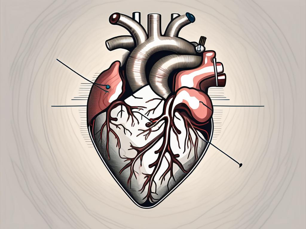
The heart produces two primary sounds, commonly referred to as S1 (the first sound) and S2 (the second sound). S1 occurs when the mitral and tricuspid valves close, while S2 occurs when the aortic and pulmonic valves close.
Let’s delve a little deeper into the intricacies of these heart sounds. S1, also known as the “lub” sound, is typically louder and longer than S2. It marks the beginning of systole, the contraction phase of the cardiac cycle. S1 is primarily caused by the closure of the mitral and tricuspid valves, which prevent blood from flowing back into the atria.
On the other hand, S2, also known as the “dub” sound, is shorter and sharper. It marks the beginning of diastole, the relaxation phase of the cardiac cycle. S2 is produced by the closure of the aortic and pulmonic valves, preventing blood from flowing back into the ventricles.
The Importance of Heart Sounds in Diagnosis
Abnormal heart sounds can indicate various cardiac conditions. Recognizing and interpreting these sounds accurately is essential for making an accurate diagnosis and providing appropriate treatment.
One common abnormality in heart sounds is the presence of an additional sound, known as S3. S3 occurs during early diastole and is often associated with conditions such as heart failure or volume overload. It is commonly referred to as the “ventricular gallop” and can be heard as a low-frequency sound following S2.
Another abnormality is the presence of a fourth heart sound, known as S4. S4 occurs just before S1 and is associated with conditions such as hypertension or ventricular hypertrophy. It is commonly referred to as the “atrial gallop” and can be heard as a low-frequency sound preceding S1.
By carefully listening to and analyzing heart sounds, healthcare professionals can gather valuable information about the condition of the heart and identify any potential abnormalities. This information, combined with other diagnostic tests, helps in formulating an accurate diagnosis and developing an appropriate treatment plan.
Tools for Auscultating Heart Sounds
To auscultate heart sounds, the primary tool you’ll need is a stethoscope. This instrument allows you to listen to the internal sounds of the body, including the heart and lungs.
The Stethoscope: Your Primary Tool
A stethoscope consists of a chest piece, tubing, and earpieces. The chest piece incorporates a diaphragm and a bell. The diaphragm is used for high-frequency sounds, such as S1 and S2, while the bell is more sensitive to low-frequency sounds like murmurs.
Let’s delve deeper into the components of a stethoscope. The diaphragm, which is the larger side of the chest piece, is a flat, circular membrane made of high-quality material that vibrates when it comes into contact with the body. This vibration allows the sound waves to travel through the tubing and into your ears, enabling you to hear the intricate details of the heart sounds.
On the other hand, the bell, which is the smaller side of the chest piece, has a concave shape and is covered with a thin layer of rubber or plastic. This design allows it to pick up low-frequency sounds with greater sensitivity. By applying gentle pressure with the bell against the patient’s chest, you can detect subtle murmurs and abnormal heart sounds that may require further investigation.
Advanced Tools for Heart Sound Auscultation
In addition to a stethoscope, some clinicians may choose to use advanced tools such as electronic stethoscopes or phonocardiograms to enhance the clarity and accuracy of auscultated sounds.
Electronic stethoscopes, for example, have the ability to amplify heart sounds, filter out background noise, and even record the audio for later analysis. This technology can be particularly useful in noisy environments or when dealing with patients who have faint heart sounds.
Phonocardiograms, on the other hand, are graphical representations of heart sounds. They provide a visual display of the different components of the cardiac cycle, allowing clinicians to analyze the timing, intensity, and characteristics of each sound in greater detail. This tool can aid in the diagnosis of various cardiac conditions, such as valve disorders or abnormal heart rhythms.
While these advanced tools can be valuable additions to a clinician’s arsenal, it’s important to note that they are not always necessary for routine heart sound auscultation. A well-trained ear and a good quality stethoscope are often sufficient to detect and interpret most cardiac abnormalities.
Preparing for Auscultation
Before starting the auscultation process, it is crucial to ensure both the patient and the environment are adequately prepared.
Patient Preparation
Instruct the patient to lie down on an examination table in a comfortable position. Ensure that the patient’s chest is exposed, as this will allow for proper placement of the stethoscope.
Once the patient is in position, it is essential to establish a sense of trust and comfort. Take a moment to explain the auscultation process to the patient, reassuring them that it is a painless and non-invasive procedure. Encourage them to ask any questions or express any concerns they may have, as this will help alleviate any anxiety they may be feeling.
Furthermore, it is important to consider the patient’s privacy and dignity during the preparation phase. Provide them with a gown or draping to cover themselves if they feel more comfortable doing so. Respecting their autonomy and ensuring their comfort will contribute to a more successful auscultation experience.
Environment and Equipment Preparation
Choose a quiet room with minimal background noise to conduct the auscultation. This will help eliminate any potential distractions that could interfere with your ability to accurately listen to the patient’s sounds. Consider turning off any electronic devices or equipment that may produce unnecessary noise.
Additionally, ensure that the lighting in the room is adequate. Proper lighting will allow you to visualize the patient’s chest and the placement of the stethoscope more effectively. Dim or harsh lighting can make it challenging to identify landmarks or potential abnormalities during the auscultation process.
Before beginning the auscultation, take a moment to inspect your stethoscope. Ensure that it is in good working condition, with no visible damage or defects. Check the tubing for any cracks or leaks that could compromise the quality of sound transmission. It is also crucial to clean the earpieces thoroughly, as any debris or buildup can affect the clarity of the sounds you hear.
When inserting the earpieces, make sure they fit securely in your ears. A proper fit will not only enhance your comfort but also prevent any extraneous noise from leaking in, allowing you to focus solely on the patient’s sounds. Adjust the tension of the earpieces if necessary, ensuring a snug yet comfortable fit.
By taking the time to adequately prepare both the patient and the environment, you are setting the stage for a successful auscultation. These preparatory steps not only contribute to accurate sound interpretation but also demonstrate your commitment to patient-centered care.
The Auscultation Process
Now that you’re ready to begin auscultating heart sounds, let’s discuss the step-by-step process to ensure accurate results.
Before we delve into the details of the auscultation process, it’s important to understand the significance of this diagnostic technique. Auscultation, derived from the Latin word “auscultare” meaning “to listen,” is a fundamental skill used by healthcare professionals to assess the health of the heart and detect any abnormalities in its functioning.
Identifying the Auscultation Points
There are specific areas on the chest where each heart sound is best heard. Familiarize yourself with these auscultation points to ensure proper placement of the stethoscope.
The four main auscultation points are:
- The aortic area, located at the second right intercostal space, where the sound of the aortic valve can be heard.
- The pulmonic area, situated at the second left intercostal space, where the sound of the pulmonic valve is best heard.
- The tricuspid area, found at the lower left sternal border, where the sound of the tricuspid valve can be auscultated.
- The mitral area, located at the fifth intercostal space in the midclavicular line, where the sound of the mitral valve is most prominent.
By accurately identifying these auscultation points, healthcare professionals can ensure that they are placing the diaphragm of the stethoscope in the optimal position to capture the heart sounds.
The Technique of Auscultation
Place the diaphragm of the stethoscope firmly over the auscultation points. Listen carefully to the sounds while taking note of the duration, intensity, and any additional sounds, such as murmurs or gallops.
During the auscultation process, it is crucial to maintain a quiet environment to minimize external noise interference. This allows for a more accurate interpretation of the heart sounds. Additionally, it is recommended to use both the diaphragm and the bell of the stethoscope to assess different frequencies of heart sounds.
As you listen to the heart sounds, pay attention to the S1 and S2 sounds, which correspond to the closure of the mitral and aortic valves, respectively. These sounds are often described as a “lub-dub” rhythm. Abnormalities in these sounds, such as a split S2 or an accentuated S1, can provide valuable information about the heart’s condition.
Furthermore, be vigilant for the presence of additional sounds, such as murmurs or gallops. Murmurs are abnormal heart sounds caused by turbulent blood flow, while gallops are extra heart sounds that can indicate underlying cardiac issues. Identifying and characterizing these additional sounds can help in diagnosing specific heart conditions.
By following these steps and honing your auscultation skills, you will be able to gather valuable information about the heart’s function and detect any potential abnormalities. Remember, practice and experience are key to becoming proficient in this essential diagnostic technique.
Interpreting Heart Sounds
Interpreting heart sounds requires knowledge of both normal and abnormal findings. Let’s dive deeper into the fascinating world of auscultation and explore the intricacies of the human heart.
The heart, a remarkable organ, beats tirelessly to pump oxygenated blood throughout the body. Auscultation, the act of listening to the heart sounds, provides valuable insights into its function and potential abnormalities.
Normal Heart Sounds
Normal heart sounds should consist of a clear S1 and S2 without any additional sounds or murmurs. S1, also known as the first heart sound or the “lub,” is caused by the closure of the mitral and tricuspid valves. Conversely, S2, the second heart sound or the “dub,” is produced by the closing of the aortic and pulmonary valves. Understanding the normal sounds is vital in distinguishing abnormal findings.
When listening to the heart, it is crucial to appreciate the subtle nuances that can affect the quality of the sounds. Factors such as body position, respiration, and patient characteristics can influence the intensity and timing of the heart sounds. For example, a forceful S1 may be heard in individuals with a hyperdynamic circulation, such as athletes or pregnant women.
Abnormal Heart Sounds
Abnormal heart sounds may include murmurs, extra heart sounds (S3 and S4), or other sounds indicating valvular or structural abnormalities. Murmurs, often described as whooshing or swishing sounds, can be indicative of turbulent blood flow caused by valve disorders or congenital defects. Extra heart sounds, such as S3 and S4, can provide valuable clues about the heart’s ability to relax and fill with blood.
Recognizing and interpreting these sounds will aid in diagnosing specific cardiac conditions. For instance, a high-pitched, blowing murmur heard during systole may suggest aortic stenosis, while a low-pitched rumbling murmur heard during diastole may indicate mitral stenosis.
Furthermore, the use of advanced auscultatory techniques, such as phonocardiography, can enhance the accuracy of heart sound interpretation. Phonocardiography involves the use of specialized sensors to capture and analyze the heart sounds, providing a visual representation of the sound waves. This technology enables healthcare professionals to identify subtle abnormalities that may not be easily discernible through traditional auscultation.
Interpreting heart sounds is a skill that requires a keen ear, a deep understanding of cardiac physiology, and a commitment to continuous learning. By honing this skill, healthcare providers can uncover vital information that guides patient care and contributes to improved cardiovascular outcomes.
Common Challenges in Auscultating Heart Sounds
While auscultating heart sounds, you may encounter certain challenges. Let’s explore some of these difficulties and how to overcome them.
Overcoming Difficulties in Hearing Heart Sounds
In some cases, external factors such as obesity or patient movement may make it challenging to hear heart sounds clearly. Experimenting with different stethoscope positions or using additional techniques such as the “bell and diaphragm” technique can help overcome these challenges.
When dealing with obesity, it’s important to consider the positioning of the patient. Sometimes, adjusting the patient’s posture or asking them to take a deep breath and hold it can help create better access to the heart sounds. Additionally, using a stethoscope with a longer tubing length can also aid in amplifying the sounds, allowing for a clearer auscultation.
Another common challenge is patient movement. Patients may unintentionally shift or fidget during the examination, causing the stethoscope to lose contact with the chest wall. To address this, it’s essential to communicate with the patient and ask them to remain as still as possible. If necessary, gently hold the stethoscope in place to maintain consistent contact with the chest.
Dealing with Patient-related Challenges
Some patients may experience anxiety, discomfort, or breathing difficulties during the auscultation process. Communicate with your patients, reassure them, and adjust the examination technique if necessary to ensure their comfort.
Anxiety can significantly affect a patient’s ability to relax, leading to increased heart rate and shallow breathing. To alleviate anxiety, create a calm and soothing environment, explain the procedure in a reassuring manner, and encourage deep breathing exercises before and during the auscultation. Taking a few moments to establish a connection with the patient and addressing any concerns they may have can also help ease their anxiety.
In cases where patients experience discomfort, it’s crucial to be attentive and responsive. If a patient reports pain or discomfort during the examination, consider adjusting the pressure applied by the stethoscope or exploring alternative positions that may provide better comfort without compromising the quality of the auscultation.
Furthermore, patients with breathing difficulties may find it challenging to maintain a steady breath while the examination is being conducted. In such cases, it may be helpful to ask the patient to take slow, deep breaths and pause briefly during the auscultation process. This allows them to catch their breath and ensures a more accurate assessment of the heart sounds.
Tips for Improving Your Auscultation Skills
Auscultation skills improve with practice and experience. Here are some tips to enhance your ability to auscultate heart sounds accurately:
Practice Strategies for Better Auscultation
Regularly practice auscultation on both healthy individuals and patients with known heart conditions. This will help you develop a discerning ear and improve your ability to identify abnormal sounds.
Continuing Education and Resources for Auscultation
Stay updated with the latest research and developments in the field of cardiology. Attend conferences, workshops, and seminars to enhance your knowledge and skills in auscultation. Additionally, refer to reputable resources, such as books, online courses, and interactive tutorials focused on heart sound auscultation.
With a thorough understanding of heart sounds, appropriate tools, and proper technique, you can become proficient in auscultating heart sounds. Mastering this skill will undoubtedly contribute to improved diagnostic accuracy and better patient care.