what sounds does a healthy heart make

When it comes to the health of our heart, one of the key indicators is the sounds it produces. Our heart creates distinct sounds, which can be heard with the aid of medical equipment such as a stethoscope or through more advanced methods like echocardiograms. Understanding these sounds and their implications is crucial for assessing cardiovascular health and identifying potential heart conditions. In this article, we will explore the basics of heart sounds, their significance, and what they can tell us about our heart’s well-being.

Understanding the Basics of Heart Sounds

Before delving into the details, it is important to grasp the essentials of heart sounds. The sounds our hearts make are a direct result of the intricate anatomy and physiology of this vital organ. By familiarizing ourselves with the anatomy of the heart and its role in producing these sounds, we can better comprehend their significance.

The Anatomy of the Heart and Its Role in Producing Sounds

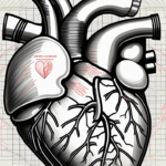
The heart is composed of four chambers – two atria and two ventricles – along with several valves that regulate blood flow. Each chamber and valve plays a crucial part in producing the sounds we hear.

The first heart sound, often referred to as the “lub” sound, is primarily caused by the closure of the mitral and tricuspid valves. This sound signifies the beginning of the cardiac cycle, when the ventricles contract and blood is pumped out of the heart.

The second heart sound, commonly known as the “dub” sound, is produced by the closure of the aortic and pulmonary valves. This sound marks the end of the cardiac cycle, as the ventricles relax and refill with blood.

The Science Behind Heart Sounds

Heart sounds, though seemingly simple, are the result of complex physiological processes. The production of these sounds involves the synchronization of muscle contractions, valve movements, and blood flow within the heart.

As blood flows through the chambers and valves, it creates specific vibrations and turbulent sounds. These vibrations, in turn, cause the audible heart sounds we perceive.

But what happens when the heart is not functioning optimally? In certain medical conditions, such as heart murmurs, the heart sounds may deviate from the normal “lub-dub” pattern. Heart murmurs are abnormal sounds that can be heard during a heartbeat and may indicate underlying heart problems.

Heart murmurs can be caused by various factors, including valve abnormalities, heart defects, or even turbulent blood flow. The identification and interpretation of heart murmurs require the expertise of healthcare professionals, such as cardiologists, who utilize specialized tools like stethoscopes to listen for these abnormal sounds.

Identifying the Sounds of a Healthy Heart

Recognizing the sounds of a healthy heart is vital for distinguishing between normal and abnormal cardiac function. By understanding what constitutes a healthy heart sound, we can better assess an individual’s cardiovascular well-being.

The “Lub-Dub” Sound Explained

A healthy heart produces a distinctive “lub-dub” sound, which is a combination of the first and second heart sounds. This rhythmic sound is strong, clear, and evenly spaced.

The “lub” sound is slightly longer and lower in pitch compared to the higher-pitched “dub” sound. Together, they create a symphony that represents the healthy functioning of the heart.

Variations in Heart Sounds and What They Mean

While the “lub-dub” sound is the norm for a healthy heart, variations in heart sounds can indicate underlying issues. It is important to be aware of these variations and understand their potential implications.

If the sounds are irregular, muffled, or accompanied by additional noises such as murmurs or clicks, it could signify valve abnormalities, a heart murmur, or other cardiac conditions that require further investigation.

Furthermore, it is worth noting that heart sounds can also vary depending on a person’s age and physical condition. For example, in children, the heart sounds may be slightly different due to their smaller heart size and thinner chest walls. Similarly, athletes may have a stronger and more pronounced “lub-dub” sound due to their well-conditioned cardiovascular system.

Additionally, certain external factors can affect heart sounds. For instance, obesity or excess body fat can make it more challenging to hear the heart sounds clearly, as the layers of fat act as a barrier. On the other hand, a thin individual may have more audible heart sounds due to the reduced tissue between the heart and the stethoscope.

The Role of Medical Equipment in Listening to Heart Sounds

Listening to heart sounds is an essential part of cardiovascular examinations. Various medical devices aid healthcare professionals in accurately assessing these sounds and interpreting their meaning.

Using a Stethoscope to Hear Heart Sounds

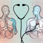
A stethoscope is the most common tool used to listen to heart sounds. It consists of a chest piece that amplifies the sounds produced by the heart and transmits them to the listener’s ears. This simple yet invaluable instrument helps clinicians gather vital information about cardiac health.

When using a stethoscope, healthcare professionals carefully place the chest piece on specific areas of the patient’s chest, known as auscultation points. These points correspond to different areas of the heart, such as the aortic, pulmonic, tricuspid, and mitral valves. By listening to the sounds produced at each auscultation point, clinicians can detect abnormalities or irregularities in the heart’s functioning.

Furthermore, stethoscopes come in various types, including acoustic and electronic models. Acoustic stethoscopes rely on the transmission of sound waves through hollow tubes, while electronic stethoscopes amplify and filter the sounds, enhancing the clinician’s ability to detect subtle nuances in the heart sounds.

Echocardiograms and Heart Sounds

In addition to stethoscopes, echocardiograms are another valuable diagnostic tool that provides comprehensive insights into heart sounds. This non-invasive imaging technique uses sound waves to create detailed images of the heart, allowing healthcare professionals to visualize its structure and function.

During an echocardiogram, a transducer is placed on the patient’s chest, emitting high-frequency sound waves that bounce off the heart’s structures. These sound waves are then converted into images, providing a real-time view of the heart’s chambers, valves, and blood flow patterns.

Echocardiograms provide valuable information about heart valve abnormalities, wall motion abnormalities, and other structural issues that may alter heart sounds. By combining the visual information obtained from an echocardiogram with the auditory data gathered through a stethoscope, healthcare professionals can form a more comprehensive understanding of a patient’s cardiovascular health.

Moreover, advancements in technology have led to the development of transesophageal echocardiograms (TEE), which involve inserting a specialized probe into the patient’s esophagus to obtain even more detailed images of the heart. TEEs are particularly useful in cases where conventional echocardiograms may not provide sufficient information.

In conclusion, medical equipment plays a crucial role in listening to heart sounds. Stethoscopes enable clinicians to detect abnormalities in the heart’s functioning, while echocardiograms provide detailed visual information about the heart’s structure and function. By utilizing these tools in conjunction, healthcare professionals can enhance their ability to diagnose and manage cardiovascular conditions effectively.

The Connection Between Heart Sounds and Heart Health

Heart sounds are closely linked to overall cardiovascular health. They serve as important indicators of the heart’s functional status and can provide valuable insights into potential heart conditions.

How Heart Sounds Indicate Cardiovascular Health

A strong, steady “lub-dub” sound suggests that the heart is functioning effectively. Regular heart sounds are an indication of normal blood flow, well-functioning valves, and healthy muscle contractions.

However, the intricacies of heart sounds go beyond just the simple “lub-dub.” Each sound has a distinct meaning and can reveal important information about the heart’s health. The first heart sound, known as S1, is caused by the closure of the mitral and tricuspid valves and signifies the beginning of systole, the contraction phase of the heart. The second heart sound, known as S2, is caused by the closure of the aortic and pulmonary valves and marks the end of systole, indicating the beginning of diastole, the relaxation phase of the heart.

In addition to S1 and S2, there are other heart sounds that can be heard under certain circumstances. For example, the third heart sound, S3, can be an early sign of heart failure. It occurs when the ventricles are filling with blood and can be heard as a low-frequency sound during diastole. The fourth heart sound, S4, is associated with stiff ventricles and can be an indicator of conditions such as hypertension or coronary artery disease.

If heart sounds deviate from their normal patterns, it could be an indication of issues such as heart valve abnormalities, heart murmurs, heart muscle disorders, or other cardiac conditions that may require medical attention.

Heart Sounds and Potential Heart Conditions

Abnormal heart sounds can provide vital clues about potential heart conditions. For instance, a heart murmur, which manifests as an abnormal sound during the cardiac cycle, may indicate valve dysfunction or a structural abnormality within the heart.

By carefully listening to and interpreting heart sounds, healthcare professionals can identify the presence of conditions such as valvular heart disease, heart failure, or congenital heart defects, guiding further diagnostic and treatment strategies.

Furthermore, the use of advanced technologies, such as echocardiography, allows for a more detailed analysis of heart sounds. This non-invasive imaging technique provides real-time visualization of the heart’s structures and allows healthcare professionals to assess the function of the valves, chambers, and overall cardiac performance. It helps in detecting abnormalities that may not be audible during a routine physical examination.

Understanding the connection between heart sounds and heart health is crucial for early detection and management of cardiovascular conditions. Regular check-ups, including thorough auscultation of the heart, can provide valuable information about the heart’s function and help ensure optimal cardiovascular health.

Frequently Asked Questions About Heart Sounds

As heart sounds can sometimes be mystifying, it is natural to have questions and misconceptions. Here, we address some common queries regarding heart sounds and provide accurate information to dispel any confusion.

Common Misconceptions About Heart Sounds

There are several misconceptions surrounding heart sounds that can lead to unnecessary worry or confusion. One common misconception is assuming that any irregularity in heart sounds is an indication of a serious heart condition.

However, it is essential to understand that the human heart is a complex organ, and its sounds can vary depending on various factors, including age, physical activity, and even emotional state. Minor variations in heart sounds can be normal and may not necessarily indicate a serious health issue.

For instance, during exercise or moments of excitement, the heart rate naturally increases, resulting in a faster rhythm and potentially louder heart sounds. These changes are usually temporary and should not cause undue concern.

When Should You Be Concerned About Heart Sounds?

Although some variations in heart sounds can be harmless, certain situations warrant immediate attention. If you experience sudden changes in heart sounds accompanied by symptoms like chest pain, dizziness, shortness of breath, or general discomfort, it is crucial to seek medical help promptly, as it could indicate an underlying cardiac emergency.

Furthermore, individuals with pre-existing heart conditions or a family history of heart disease should be particularly vigilant about any changes in heart sounds. Regular check-ups and consultations with a healthcare professional are essential to monitor and manage cardiovascular health effectively.

In summary, understanding the sounds a healthy heart makes is a valuable tool in assessing cardiovascular health. By familiarizing ourselves with the basics of heart sounds, identifying their variations, and recognizing the connection between heart sounds and heart health, we can empower ourselves to prioritize our cardiovascular well-being.

Remember, knowledge is power, but it is equally important to know when to seek professional advice. Should any doubts or concerns arise, consulting a healthcare professional is always the best course of action to gain proper insight and care for our hearts.

where to assess heart sounds

Assessing heart sounds is a crucial component of a comprehensive cardiovascular examination. By carefully listening to the heart, healthcare practitioners can gain invaluable insights into a patient’s cardiac health. Understanding where to assess heart sounds is essential for accurate and thorough evaluation. In this article, we will explore the various areas on the chest where heart sounds can be heard and the significance of each location.

Understanding Heart Sounds

The Basics of Heart Sounds

Heart sounds, also known as cardiac sounds, are the noises produced by the functioning of the heart valves and the flow of blood through the chambers. The normal heart sounds are commonly referred to as S1 and S2, representing the closure of the atrioventricular (AV) valves and semilunar valves, respectively.

S1, the first heart sound, is typically described as a “lub” sound and corresponds to the closure of the mitral and tricuspid valves. S2, the second heart sound, is often characterized as a “dub” sound and occurs when the aortic and pulmonic valves shut.

Did you know that heart sounds can vary depending on a person’s age and overall health? In addition to the S1 and S2 sounds, there are other heart sounds that can be heard under certain circumstances. For example, the third heart sound, S3, can be heard in some individuals with heart failure. This sound is often described as a “gallop” and is caused by the rapid filling of the ventricles during diastole. Similarly, the fourth heart sound, S4, can be heard in individuals with stiff ventricles, such as those with hypertension or coronary artery disease.

The Importance of Heart Sound Assessment

Assessing heart sounds can assist clinicians in detecting abnormalities and diagnosing cardiac conditions. Abnormal heart sounds, known as heart murmurs, may indicate valve disorders, such as mitral regurgitation or aortic stenosis, among other conditions. By recognizing abnormal heart sounds, healthcare professionals can promptly intervene and provide appropriate treatment.

It is important to note that heart sound assessment is not only useful for diagnosing cardiac conditions but also for monitoring the progress of treatment. For example, a decrease in the intensity of a heart murmur may indicate that a valve repair or replacement surgery was successful. Conversely, the appearance of new or worsening heart sounds may suggest a complication or the need for further intervention.

Next time you hear your heart beating, take a moment to appreciate the intricate sounds it produces. Each lub and dub is a testament to the complex workings of this vital organ. And remember, by understanding heart sounds, we can better understand and care for our cardiovascular health.

Anatomy of the Heart and Sound Production

The Heart’s Structure and Function

Before we explore where to assess heart sounds, it is crucial to have a basic understanding of the heart’s structure and function. The heart is a hollow, muscular organ located in the chest. It consists of four chambers: the left and right atria and the left and right ventricles. The atria receive blood, while the ventricles pump blood out of the heart.

Within the heart, there is a complex network of blood vessels that supply oxygen and nutrients to the heart muscle itself, known as the coronary arteries. These arteries ensure that the heart receives the necessary nourishment to continue beating and pumping blood throughout the body.

The heart is also equipped with a specialized electrical conduction system that coordinates the rhythmic contractions of its chambers. This system includes the sinoatrial (SA) node, which acts as the heart’s natural pacemaker, initiating the electrical impulses that regulate the heart rate. The impulses then travel through the atria, causing them to contract, and eventually reach the ventricles, leading to their contraction as well.

How Heart Sounds are Produced

The closure of heart valves during the cardiac cycle produces the distinctive heart sounds. The S1 sound is created when the mitral and tricuspid valves shut, and blood is forced into the ventricles. This sound is often described as a “lub” and marks the beginning of systole, the phase of the cardiac cycle when the heart contracts and pumps blood out of the ventricles.

Following the S1 sound, there is a brief period of silence as the ventricles fill with blood. This is known as the isovolumetric relaxation phase. As the ventricles continue to fill, the pressure within them rises until it exceeds the pressure in the aorta and pulmonary artery. At this point, the aortic and pulmonic valves open, allowing blood to be ejected from the heart.

The S2 sound occurs when the aortic and pulmonic valves close as blood is ejected from the heart to the rest of the body. This sound, often described as a “dub,” marks the beginning of diastole, the phase of the cardiac cycle when the heart relaxes and refills with blood.

Understanding the anatomy of the heart and the production of heart sounds is essential for healthcare professionals to accurately assess and interpret these sounds. By listening carefully to the timing, intensity, and quality of heart sounds, clinicians can gather valuable information about the functioning of the heart and detect any abnormalities that may require further investigation or treatment.

Techniques for Assessing Heart Sounds

Auscultation Method

Auscultation, or listening with a stethoscope, is the primary method to assess heart sounds. To perform auscultation, healthcare practitioners place the stethoscope diaphragm or bell on specific areas of the chest to listen to the heart sounds. This technique allows practitioners to isolate and evaluate each valve area separately.

During auscultation, healthcare practitioners pay close attention to the timing, intensity, and quality of the heart sounds. The timing of the heart sounds can provide valuable information about the cardiac cycle and the functioning of the heart. For example, the first heart sound (S1) is typically heard as the mitral and tricuspid valves close, marking the beginning of systole. The second heart sound (S2) is heard as the aortic and pulmonic valves close, marking the end of systole. Any abnormalities in the timing of these sounds can indicate underlying cardiac conditions.

Furthermore, the intensity of the heart sounds can vary and may provide additional clues about the heart’s health. For instance, a loud S1 sound may suggest mitral stenosis, while a soft S2 sound may indicate aortic regurgitation. By carefully assessing the intensity of the heart sounds, healthcare practitioners can gather important information to aid in diagnosis and treatment decisions.

Palpation Method

In addition to auscultation, palpation can provide valuable information during heart sound assessment. Palpation involves gently placing the fingers over the pulsation points on the chest to feel for abnormal vibrations or thrills, which may indicate underlying pathology.

During palpation, healthcare practitioners assess the strength and regularity of the heartbeat. They may feel for any abnormal pulsations or thrills, which can be indicative of conditions such as heart murmurs or abnormal blood flow. By carefully palpating the chest, healthcare practitioners can gather additional information to complement their auscultation findings.

Moreover, palpation can also help healthcare practitioners assess the peripheral pulses. By comparing the strength and regularity of the peripheral pulses to the heart sounds, practitioners can gain a more comprehensive understanding of the cardiovascular system’s overall function. This information can be particularly useful in diagnosing conditions such as peripheral artery disease or assessing the effectiveness of certain treatments.

Locations to Assess Heart Sounds

The human heart is a remarkable organ that pumps blood throughout the body, ensuring the delivery of oxygen and nutrients to every cell. To assess the function of the heart, healthcare professionals rely on auscultation, the act of listening to heart sounds using a stethoscope. By carefully placing the stethoscope at specific locations on the chest, they can gather valuable information about the heart’s performance.

One of the key locations for assessing heart sounds is the aortic area. This area, also known as the second right intercostal space, is where the aortic valve sounds can be heard. To locate this area, find the second rib on the right side of the chest, and move your stethoscope slightly to the right. Here, you will primarily hear the first and second heart sounds (S1 and S2) associated with the aortic valve.

Another important location for assessing heart sounds is the pulmonic area. This area is found at the second left intercostal space. To access this area, move your stethoscope slightly to the left from the second rib. By focusing your attention here, you can listen to the pulmonic valve sounds, including S1 and S2. These sounds provide insights into the functioning of the pulmonary valve and the flow of blood to the lungs.

Next, we have the tricuspid area, which is located at the left lower sternal border, near the fifth intercostal space. Placing your stethoscope in this area allows you to hear the tricuspid valve sounds, including S1 and S2. These sounds are crucial in evaluating the function of the tricuspid valve, which separates the right atrium from the right ventricle.

Lastly, we have the mitral area, also known as the cardiac apex. This area is located at the fifth intercostal space and the midclavicular line on the left side of the chest. Positioning your stethoscope at this spot enables you to listen to the mitral valve sounds, including S1 and S2. These sounds provide valuable information about the function of the mitral valve, which separates the left atrium from the left ventricle.

By carefully assessing heart sounds at these specific locations, healthcare professionals can detect abnormalities, such as murmurs or irregular rhythms, which may indicate underlying heart conditions. It is through their expertise and attention to detail that they can provide accurate diagnoses and develop appropriate treatment plans to ensure the well-being of their patients.

Interpreting Heart Sounds

Normal Heart Sounds

Understanding the characteristics of normal heart sounds is crucial for accurate interpretation. A clear and distinct “lub-dub” rhythm signifies the normal closure of the heart valves. This rhythmic sound is produced by the coordinated contraction and relaxation of the heart chambers. The first heart sound, or S1, is caused by the closure of the mitral and tricuspid valves, while the second heart sound, or S2, is produced by the closure of the aortic and pulmonary valves.

During S1, the mitral and tricuspid valves snap shut, creating a low-pitched sound that is best heard at the apex of the heart. This sound marks the beginning of systole, the phase when the heart contracts and pumps blood out to the body. Following S1, there is a brief pause as the heart chambers fill with blood.

As the heart relaxes during diastole, the aortic and pulmonary valves close, producing the second heart sound, S2. This sound is higher-pitched and is best heard at the base of the heart. S2 marks the end of systole and the beginning of diastole, when the heart chambers fill with blood again.

Abnormal Heart Sounds

Abnormal heart sounds, known as heart murmurs, may suggest structural issues with the heart valves or blood flow restrictions. Heart murmurs can be systolic, diastolic, or continuous in nature. Systolic murmurs occur between S1 and S2, while diastolic murmurs occur between S2 and the next S1. Continuous murmurs are heard throughout the cardiac cycle.

Identifying the location, timing, and intensity of the murmurs allows healthcare professionals to determine the potential causes accurately. For example, a systolic murmur heard best at the apex of the heart may indicate mitral valve regurgitation, where blood leaks backward through the valve during systole. On the other hand, a diastolic murmur heard best at the left sternal border may suggest aortic valve stenosis, a condition characterized by the narrowing of the aortic valve opening.

It is important to note that not all heart murmurs are pathological. Innocent or functional murmurs can occur in individuals without any structural heart abnormalities. These murmurs are usually benign and are commonly heard in children and young adults during periods of rapid growth or increased blood flow.

The Role of Technology in Heart Sound Assessment

Electronic Stethoscopes

Technological advancements have introduced electronic stethoscopes that enhance heart sound assessment. These devices amplify the heart sounds, allowing for more precise and detailed evaluation. Some electronic stethoscopes also provide visual displays, enabling clinicians to observe the waveforms and detect subtle abnormalities.

Echocardiograms

Echocardiograms, or cardiac ultrasound examinations, play a significant role in assessing heart sounds. This non-invasive imaging technique utilizes sound waves to create detailed images of the heart’s structure, function, and blood flow. Echocardiograms provide valuable information about cardiac anatomy and assist in diagnosing conditions such as valve disorders, congenital defects, and heart failure.

Cardiac Magnetic Resonance Imaging (MRI)

In addition to electronic stethoscopes and echocardiograms, cardiac magnetic resonance imaging (MRI) is another technology that aids in heart sound assessment. This imaging technique uses a powerful magnetic field and radio waves to create detailed images of the heart. Cardiac MRI provides information about the heart’s size, shape, and function, allowing clinicians to assess heart sounds with greater accuracy.

Artificial Intelligence (AI) Applications

Advancements in artificial intelligence (AI) have also contributed to the field of heart sound assessment. AI algorithms can analyze heart sounds and identify patterns or abnormalities that may not be easily detectable by human ears. This technology assists clinicians in making more accurate diagnoses and treatment decisions, improving patient outcomes.

Telemedicine and Remote Monitoring

With the rise of telemedicine, technology has enabled heart sound assessment to be conducted remotely. Patients can record their heart sounds using electronic stethoscopes or smartphone applications and transmit the recordings to healthcare providers for evaluation. This allows for continuous monitoring and early detection of any changes or abnormalities in heart sounds, leading to timely intervention and improved patient care.

Overall, technology has revolutionized heart sound assessment, providing clinicians with advanced tools and techniques to accurately evaluate and diagnose various cardiac conditions. From electronic stethoscopes and echocardiograms to cardiac MRI and AI applications, these technological advancements have significantly enhanced our understanding of heart sounds and improved patient care.

Common Challenges in Assessing Heart Sounds

Patient Factors

Assessing heart sounds can be challenging due to various patient factors. Obesity, chest deformities, and the presence of lung disease can make it difficult to obtain clear heart sounds. Additionally, patient anxiety or physical discomfort may affect the accuracy of the assessment. In such cases, repeated attempts or alternative assessment methods may be necessary.

Practitioner Skill Level

Accurately interpreting heart sounds requires skill and experience. Novice practitioners may face challenges in differentiating normal heart sounds from abnormalities. Continuous learning, practice, and mentorship are essential to improving diagnostic capabilities and ensuring accurate heart sound assessment.

Tips for Accurate Heart Sound Assessment

Patient Preparation

To facilitate accurate heart sound assessment, proper patient preparation is crucial. Ensure a comfortable and relaxed environment for the patient, explaining the procedure and anticipated sensations. If necessary, position the patient appropriately to optimize access to the cardiac areas.

Optimal Environment for Assessment

Conducting heart sound assessment in a quiet room minimizes external noise interference. Reduce distractions, such as electronic devices or unnecessary conversations, to focus on the sounds emanating from the heart.

Continuous Learning and Practice

Heart sound assessment is a skill that improves with frequent practice and continuous education. Medical practitioners should actively seek opportunities to enhance their knowledge and hands-on experience in order to master this critical aspect of cardiovascular evaluation.

In summary, knowing where to assess heart sounds is essential for accurate diagnosis and management of various cardiac conditions. By understanding the basics of heart sounds, the anatomical locations for assessment, and the role of technology in sound interpretation, healthcare professionals can provide comprehensive care and improve patient outcomes. Overcoming challenges and continually striving for accuracy through practice and education ensures that heart sound assessment remains a vital tool in cardiovascular examination.

how to auscultate heart sounds

Auscultating heart sounds is an essential skill for healthcare professionals. By listening carefully to the sounds produced by the heart, clinicians can identify potential abnormalities and diagnose various cardiac conditions. In this article, we will explore the process of auscultating heart sounds, the tools required, and tips for improving your auscultation skills.

Understanding Heart Sounds

Heart sounds provide valuable information about the functioning of the heart. It is crucial to have a firm understanding of the basics of heart sounds before diving into the auscultation process.

The Basics of Heart Sounds

The heart produces two primary sounds, commonly referred to as S1 (the first sound) and S2 (the second sound). S1 occurs when the mitral and tricuspid valves close, while S2 occurs when the aortic and pulmonic valves close.

Let’s delve a little deeper into the intricacies of these heart sounds. S1, also known as the “lub” sound, is typically louder and longer than S2. It marks the beginning of systole, the contraction phase of the cardiac cycle. S1 is primarily caused by the closure of the mitral and tricuspid valves, which prevent blood from flowing back into the atria.

On the other hand, S2, also known as the “dub” sound, is shorter and sharper. It marks the beginning of diastole, the relaxation phase of the cardiac cycle. S2 is produced by the closure of the aortic and pulmonic valves, preventing blood from flowing back into the ventricles.

The Importance of Heart Sounds in Diagnosis

Abnormal heart sounds can indicate various cardiac conditions. Recognizing and interpreting these sounds accurately is essential for making an accurate diagnosis and providing appropriate treatment.

One common abnormality in heart sounds is the presence of an additional sound, known as S3. S3 occurs during early diastole and is often associated with conditions such as heart failure or volume overload. It is commonly referred to as the “ventricular gallop” and can be heard as a low-frequency sound following S2.

Another abnormality is the presence of a fourth heart sound, known as S4. S4 occurs just before S1 and is associated with conditions such as hypertension or ventricular hypertrophy. It is commonly referred to as the “atrial gallop” and can be heard as a low-frequency sound preceding S1.

By carefully listening to and analyzing heart sounds, healthcare professionals can gather valuable information about the condition of the heart and identify any potential abnormalities. This information, combined with other diagnostic tests, helps in formulating an accurate diagnosis and developing an appropriate treatment plan.

Tools for Auscultating Heart Sounds

To auscultate heart sounds, the primary tool you’ll need is a stethoscope. This instrument allows you to listen to the internal sounds of the body, including the heart and lungs.

The Stethoscope: Your Primary Tool

A stethoscope consists of a chest piece, tubing, and earpieces. The chest piece incorporates a diaphragm and a bell. The diaphragm is used for high-frequency sounds, such as S1 and S2, while the bell is more sensitive to low-frequency sounds like murmurs.

Let’s delve deeper into the components of a stethoscope. The diaphragm, which is the larger side of the chest piece, is a flat, circular membrane made of high-quality material that vibrates when it comes into contact with the body. This vibration allows the sound waves to travel through the tubing and into your ears, enabling you to hear the intricate details of the heart sounds.

On the other hand, the bell, which is the smaller side of the chest piece, has a concave shape and is covered with a thin layer of rubber or plastic. This design allows it to pick up low-frequency sounds with greater sensitivity. By applying gentle pressure with the bell against the patient’s chest, you can detect subtle murmurs and abnormal heart sounds that may require further investigation.

Advanced Tools for Heart Sound Auscultation

In addition to a stethoscope, some clinicians may choose to use advanced tools such as electronic stethoscopes or phonocardiograms to enhance the clarity and accuracy of auscultated sounds.

Electronic stethoscopes, for example, have the ability to amplify heart sounds, filter out background noise, and even record the audio for later analysis. This technology can be particularly useful in noisy environments or when dealing with patients who have faint heart sounds.

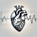
Phonocardiograms, on the other hand, are graphical representations of heart sounds. They provide a visual display of the different components of the cardiac cycle, allowing clinicians to analyze the timing, intensity, and characteristics of each sound in greater detail. This tool can aid in the diagnosis of various cardiac conditions, such as valve disorders or abnormal heart rhythms.

While these advanced tools can be valuable additions to a clinician’s arsenal, it’s important to note that they are not always necessary for routine heart sound auscultation. A well-trained ear and a good quality stethoscope are often sufficient to detect and interpret most cardiac abnormalities.

Preparing for Auscultation

Before starting the auscultation process, it is crucial to ensure both the patient and the environment are adequately prepared.

Patient Preparation

Instruct the patient to lie down on an examination table in a comfortable position. Ensure that the patient’s chest is exposed, as this will allow for proper placement of the stethoscope.

Once the patient is in position, it is essential to establish a sense of trust and comfort. Take a moment to explain the auscultation process to the patient, reassuring them that it is a painless and non-invasive procedure. Encourage them to ask any questions or express any concerns they may have, as this will help alleviate any anxiety they may be feeling.

Furthermore, it is important to consider the patient’s privacy and dignity during the preparation phase. Provide them with a gown or draping to cover themselves if they feel more comfortable doing so. Respecting their autonomy and ensuring their comfort will contribute to a more successful auscultation experience.

Environment and Equipment Preparation

Choose a quiet room with minimal background noise to conduct the auscultation. This will help eliminate any potential distractions that could interfere with your ability to accurately listen to the patient’s sounds. Consider turning off any electronic devices or equipment that may produce unnecessary noise.

Additionally, ensure that the lighting in the room is adequate. Proper lighting will allow you to visualize the patient’s chest and the placement of the stethoscope more effectively. Dim or harsh lighting can make it challenging to identify landmarks or potential abnormalities during the auscultation process.

Before beginning the auscultation, take a moment to inspect your stethoscope. Ensure that it is in good working condition, with no visible damage or defects. Check the tubing for any cracks or leaks that could compromise the quality of sound transmission. It is also crucial to clean the earpieces thoroughly, as any debris or buildup can affect the clarity of the sounds you hear.

When inserting the earpieces, make sure they fit securely in your ears. A proper fit will not only enhance your comfort but also prevent any extraneous noise from leaking in, allowing you to focus solely on the patient’s sounds. Adjust the tension of the earpieces if necessary, ensuring a snug yet comfortable fit.

By taking the time to adequately prepare both the patient and the environment, you are setting the stage for a successful auscultation. These preparatory steps not only contribute to accurate sound interpretation but also demonstrate your commitment to patient-centered care.

The Auscultation Process

Now that you’re ready to begin auscultating heart sounds, let’s discuss the step-by-step process to ensure accurate results.

Before we delve into the details of the auscultation process, it’s important to understand the significance of this diagnostic technique. Auscultation, derived from the Latin word “auscultare” meaning “to listen,” is a fundamental skill used by healthcare professionals to assess the health of the heart and detect any abnormalities in its functioning.

Identifying the Auscultation Points

There are specific areas on the chest where each heart sound is best heard. Familiarize yourself with these auscultation points to ensure proper placement of the stethoscope.

The four main auscultation points are:

  1. The aortic area, located at the second right intercostal space, where the sound of the aortic valve can be heard.
  2. The pulmonic area, situated at the second left intercostal space, where the sound of the pulmonic valve is best heard.
  3. The tricuspid area, found at the lower left sternal border, where the sound of the tricuspid valve can be auscultated.
  4. The mitral area, located at the fifth intercostal space in the midclavicular line, where the sound of the mitral valve is most prominent.

By accurately identifying these auscultation points, healthcare professionals can ensure that they are placing the diaphragm of the stethoscope in the optimal position to capture the heart sounds.

The Technique of Auscultation

Place the diaphragm of the stethoscope firmly over the auscultation points. Listen carefully to the sounds while taking note of the duration, intensity, and any additional sounds, such as murmurs or gallops.

During the auscultation process, it is crucial to maintain a quiet environment to minimize external noise interference. This allows for a more accurate interpretation of the heart sounds. Additionally, it is recommended to use both the diaphragm and the bell of the stethoscope to assess different frequencies of heart sounds.

As you listen to the heart sounds, pay attention to the S1 and S2 sounds, which correspond to the closure of the mitral and aortic valves, respectively. These sounds are often described as a “lub-dub” rhythm. Abnormalities in these sounds, such as a split S2 or an accentuated S1, can provide valuable information about the heart’s condition.

Furthermore, be vigilant for the presence of additional sounds, such as murmurs or gallops. Murmurs are abnormal heart sounds caused by turbulent blood flow, while gallops are extra heart sounds that can indicate underlying cardiac issues. Identifying and characterizing these additional sounds can help in diagnosing specific heart conditions.

By following these steps and honing your auscultation skills, you will be able to gather valuable information about the heart’s function and detect any potential abnormalities. Remember, practice and experience are key to becoming proficient in this essential diagnostic technique.

Interpreting Heart Sounds

Interpreting heart sounds requires knowledge of both normal and abnormal findings. Let’s dive deeper into the fascinating world of auscultation and explore the intricacies of the human heart.

The heart, a remarkable organ, beats tirelessly to pump oxygenated blood throughout the body. Auscultation, the act of listening to the heart sounds, provides valuable insights into its function and potential abnormalities.

Normal Heart Sounds

Normal heart sounds should consist of a clear S1 and S2 without any additional sounds or murmurs. S1, also known as the first heart sound or the “lub,” is caused by the closure of the mitral and tricuspid valves. Conversely, S2, the second heart sound or the “dub,” is produced by the closing of the aortic and pulmonary valves. Understanding the normal sounds is vital in distinguishing abnormal findings.

When listening to the heart, it is crucial to appreciate the subtle nuances that can affect the quality of the sounds. Factors such as body position, respiration, and patient characteristics can influence the intensity and timing of the heart sounds. For example, a forceful S1 may be heard in individuals with a hyperdynamic circulation, such as athletes or pregnant women.

Abnormal Heart Sounds

Abnormal heart sounds may include murmurs, extra heart sounds (S3 and S4), or other sounds indicating valvular or structural abnormalities. Murmurs, often described as whooshing or swishing sounds, can be indicative of turbulent blood flow caused by valve disorders or congenital defects. Extra heart sounds, such as S3 and S4, can provide valuable clues about the heart’s ability to relax and fill with blood.

Recognizing and interpreting these sounds will aid in diagnosing specific cardiac conditions. For instance, a high-pitched, blowing murmur heard during systole may suggest aortic stenosis, while a low-pitched rumbling murmur heard during diastole may indicate mitral stenosis.

Furthermore, the use of advanced auscultatory techniques, such as phonocardiography, can enhance the accuracy of heart sound interpretation. Phonocardiography involves the use of specialized sensors to capture and analyze the heart sounds, providing a visual representation of the sound waves. This technology enables healthcare professionals to identify subtle abnormalities that may not be easily discernible through traditional auscultation.

Interpreting heart sounds is a skill that requires a keen ear, a deep understanding of cardiac physiology, and a commitment to continuous learning. By honing this skill, healthcare providers can uncover vital information that guides patient care and contributes to improved cardiovascular outcomes.

Common Challenges in Auscultating Heart Sounds

While auscultating heart sounds, you may encounter certain challenges. Let’s explore some of these difficulties and how to overcome them.

Overcoming Difficulties in Hearing Heart Sounds

In some cases, external factors such as obesity or patient movement may make it challenging to hear heart sounds clearly. Experimenting with different stethoscope positions or using additional techniques such as the “bell and diaphragm” technique can help overcome these challenges.

When dealing with obesity, it’s important to consider the positioning of the patient. Sometimes, adjusting the patient’s posture or asking them to take a deep breath and hold it can help create better access to the heart sounds. Additionally, using a stethoscope with a longer tubing length can also aid in amplifying the sounds, allowing for a clearer auscultation.

Another common challenge is patient movement. Patients may unintentionally shift or fidget during the examination, causing the stethoscope to lose contact with the chest wall. To address this, it’s essential to communicate with the patient and ask them to remain as still as possible. If necessary, gently hold the stethoscope in place to maintain consistent contact with the chest.

Dealing with Patient-related Challenges

Some patients may experience anxiety, discomfort, or breathing difficulties during the auscultation process. Communicate with your patients, reassure them, and adjust the examination technique if necessary to ensure their comfort.

Anxiety can significantly affect a patient’s ability to relax, leading to increased heart rate and shallow breathing. To alleviate anxiety, create a calm and soothing environment, explain the procedure in a reassuring manner, and encourage deep breathing exercises before and during the auscultation. Taking a few moments to establish a connection with the patient and addressing any concerns they may have can also help ease their anxiety.

In cases where patients experience discomfort, it’s crucial to be attentive and responsive. If a patient reports pain or discomfort during the examination, consider adjusting the pressure applied by the stethoscope or exploring alternative positions that may provide better comfort without compromising the quality of the auscultation.

Furthermore, patients with breathing difficulties may find it challenging to maintain a steady breath while the examination is being conducted. In such cases, it may be helpful to ask the patient to take slow, deep breaths and pause briefly during the auscultation process. This allows them to catch their breath and ensures a more accurate assessment of the heart sounds.

Tips for Improving Your Auscultation Skills

Auscultation skills improve with practice and experience. Here are some tips to enhance your ability to auscultate heart sounds accurately:

Practice Strategies for Better Auscultation

Regularly practice auscultation on both healthy individuals and patients with known heart conditions. This will help you develop a discerning ear and improve your ability to identify abnormal sounds.

Continuing Education and Resources for Auscultation

Stay updated with the latest research and developments in the field of cardiology. Attend conferences, workshops, and seminars to enhance your knowledge and skills in auscultation. Additionally, refer to reputable resources, such as books, online courses, and interactive tutorials focused on heart sound auscultation.

With a thorough understanding of heart sounds, appropriate tools, and proper technique, you can become proficient in auscultating heart sounds. Mastering this skill will undoubtedly contribute to improved diagnostic accuracy and better patient care.

what causes heart beat sounds

Heart beat sounds are a fundamental part of our body’s rhythm and an indicator of our cardiovascular health. Understanding the causes and significance of these sounds can provide valuable insights into the functioning of our heart. In this article, we will explore the anatomy of the heart, the science behind heart beat sounds, the different types of heart sounds, factors that can affect these sounds, and how heart conditions can be diagnosed through sound analysis. Additionally, we will discuss the importance of maintaining a healthy heart through lifestyle changes and regular check-ups.

Understanding the Anatomy of the Heart

The heart plays a crucial role in the circulatory system, which transports oxygen and nutrients throughout the body. It is composed of various components, each with its own unique function.

The Role of the Heart in the Circulatory System

The heart acts as a pump, propelling blood through a network of blood vessels. This continuous flow ensures oxygenated blood reaches all organs and tissues, while deoxygenated blood returns to the lungs for reoxygenation.

Components of the Heart and Their Functions

The heart consists of four chambers: two atria and two ventricles. The atria receive blood, while the ventricles pump it out. Valves within the heart prevent backflow, ensuring a unidirectional flow of blood.

Let’s delve deeper into the components of the heart. The atria, also known as the receiving chambers, are responsible for collecting blood from various parts of the body. Once the atria have received the blood, they contract, pushing it into the ventricles. This coordinated contraction ensures a smooth flow of blood through the heart.

Now, let’s focus on the ventricles, the pumping chambers of the heart. The left ventricle, often referred to as the powerhouse of the heart, is responsible for pumping oxygenated blood to the rest of the body. Its muscular walls are thicker than those of the right ventricle, enabling it to generate enough force to propel blood through the systemic circulation.

On the other hand, the right ventricle is responsible for pumping deoxygenated blood to the lungs for oxygenation. Although it doesn’t require as much force as the left ventricle, it still plays a vital role in the overall function of the heart.

Now that we have explored the chambers of the heart, let’s discuss the valves that ensure the unidirectional flow of blood. The tricuspid valve, located between the right atrium and right ventricle, prevents the backflow of blood into the atrium when the ventricle contracts. Similarly, the mitral valve, situated between the left atrium and left ventricle, performs the same function on the left side of the heart.

In addition to the atrioventricular valves, there are also the semilunar valves. The pulmonary valve, located between the right ventricle and the pulmonary artery, prevents blood from flowing back into the ventricle after it has been pumped out. Similarly, the aortic valve, situated between the left ventricle and the aorta, ensures that blood doesn’t flow back into the ventricle once it has been ejected.

Understanding the intricate components and functions of the heart allows us to appreciate the remarkable efficiency of this vital organ. Its ability to continuously pump blood throughout the body ensures the delivery of oxygen and nutrients to every cell, enabling us to thrive and live our lives to the fullest.

The Science Behind Heart Beat Sounds

Heart beat sounds are created during the cardiac cycle, a rhythmic contraction and relaxation of the heart muscle. This cycle is responsible for the familiar lub-dub sounds heard through a stethoscope.

The Cardiac Cycle: A Closer Look

During the cardiac cycle, the heart contracts (systole) and then relaxes (diastole). These actions result in the opening and closing of heart valves, causing the characteristic sounds.

The Role of Valves in Creating Heart Sounds

Heart sounds are produced when the heart valves close, generating vibrations in the blood and surrounding tissues. The first heart sound (S1) occurs when the atrioventricular valves close, while the second heart sound (S2) is produced by the closure of the semilunar valves.

Let’s delve deeper into the fascinating world of heart sounds. Did you know that there are additional heart sounds beyond the familiar lub-dub? These additional sounds, known as murmurs, can provide valuable information about the health of the heart.

Murmurs are abnormal heart sounds that occur due to turbulent blood flow within the heart. They can be caused by various factors, such as valve abnormalities, heart defects, or even certain medical conditions. By carefully listening to these murmurs, healthcare professionals can gather important clues about the functioning of the heart and identify potential issues.

Furthermore, the intensity, timing, and location of heart sounds can also provide valuable insights. For example, a loud and prolonged S1 sound may indicate a problem with the atrioventricular valves, while a soft or absent S2 sound could be a sign of semilunar valve dysfunction. By analyzing these subtle variations in heart sounds, healthcare professionals can make accurate diagnoses and tailor treatment plans accordingly.

Different Types of Heart Sounds

Heart sounds can vary depending on whether they are normal or abnormal. Recognizing these sounds can aid in the identification of potential heart conditions.

Understanding the different types of heart sounds is crucial in diagnosing and treating various cardiac conditions. Let’s delve deeper into the normal and abnormal heart sounds to gain a comprehensive understanding.

Normal Heart Sounds: S1 and S2

S1 and S2 are the typical sounds heard during a healthy heart’s functioning. S1, the first sound, is a low-pitched “lub” heard at the beginning of the cardiac cycle. This sound is produced by the closure of the mitral and tricuspid valves, which prevents the backflow of blood into the atria. It signifies the beginning of ventricular systole, the contraction phase of the heart.

On the other hand, S2, the second sound, is a higher-pitched “dub” occurring at the end of the cycle. It is caused by the closure of the aortic and pulmonary valves, preventing the backflow of blood into the ventricles. This sound marks the beginning of ventricular diastole, the relaxation phase of the heart.

Abnormal Heart Sounds: Murmurs and Clicks

While normal heart sounds indicate a healthy heart, abnormal heart sounds can be indicative of underlying heart problems. Murmurs, for instance, are additional sounds caused by turbulent blood flow within the heart. These sounds can be heard as a swishing or whooshing noise and can be classified into systolic or diastolic murmurs, depending on when they occur during the cardiac cycle.

Clicks, on the other hand, are sharp sounds associated with valve problems or structural abnormalities. They are often caused by the opening or closing of abnormal heart valves. These abnormal sounds can be indicative of conditions such as mitral valve prolapse or aortic stenosis.

It is important to note that not all murmurs or clicks are necessarily harmful or require immediate medical attention. Some individuals may have innocent or functional murmurs, which are harmless and do not indicate any underlying heart conditions. However, it is always recommended to consult a healthcare professional for a thorough evaluation if you experience any abnormal heart sounds.

Factors Affecting the Heart Beat Sounds

Heart beat sounds can be influenced by various factors, including age and certain health conditions. Understanding these factors can provide valuable insights into the complexities of the human cardiovascular system.

Age and Heart Sounds

As individuals age, changes occur within the heart that can affect the intensity and clarity of heart sounds. The heart, like any other organ, undergoes wear and tear over time. This natural aging process can lead to the deterioration of heart valves and the hardening of blood vessels.

Imagine the heart valves as tiny gatekeepers, ensuring the smooth flow of blood through the chambers. Over the years, these valves may become less flexible and efficient, causing turbulence in the blood flow. As a result, the heart sounds may become fainter or less distinct, making it more challenging for healthcare professionals to interpret them accurately.

Health Conditions and Their Impact on Heart Sounds

Certain health conditions can have a profound impact on heart beat sounds. Heart valve abnormalities, for instance, can disrupt the normal flow of blood, leading to changes in heart sounds. These abnormalities can manifest as murmurs, which are abnormal sounds heard during the heartbeat cycle.

Structural defects within the heart can also contribute to alterations in heart beat sounds. These defects may include congenital malformations or acquired conditions that affect the heart’s anatomy. In such cases, additional sounds may be present during the cardiac cycle, indicating the need for further medical investigation.

It is crucial to recognize that changes in heart beat sounds can serve as valuable diagnostic clues for healthcare professionals. By carefully listening to and analyzing these sounds, medical experts can detect underlying conditions and provide appropriate treatment. Therefore, understanding the factors that affect heart sounds is essential in the field of cardiology, enabling healthcare providers to deliver accurate diagnoses and effective interventions.

Diagnosing Heart Conditions Through Sounds

Heart beat sounds are valuable diagnostic tools that can assist healthcare professionals in determining the presence of heart conditions. The ability to listen to the heart sounds is a skill that has been honed over centuries, and it continues to be an essential part of a thorough physical examination.

A stethoscope is commonly used to listen to heart sounds during a physical examination. This simple yet invaluable tool enables healthcare providers to detect abnormalities, such as murmurs or irregularities in heart rhythm, which may warrant further investigation. The stethoscope consists of a chest piece, which is placed on the patient’s chest, and earpieces, which allow the healthcare provider to listen to the sounds produced by the heart.

The Use of Stethoscope in Listening to Heart Sounds

When the stethoscope is placed on the chest, the healthcare provider can hear the distinct sounds produced by the heart. These sounds are created by the opening and closing of the heart valves, as well as the flow of blood through the chambers of the heart. By carefully listening to these sounds, healthcare professionals can gather valuable information about the condition of the heart.

During the examination, the healthcare provider listens for specific sounds, such as the lub-dub sound, which is the normal sound of a healthy heart. Any deviations from this normal sound can indicate the presence of a heart condition. For example, a murmur, which is an abnormal sound caused by turbulent blood flow, may suggest a problem with the heart valves.

Modern Techniques in Heart Sound Analysis

Advancements in technology have led to the development of innovative techniques, such as phonocardiography and computer-assisted analyses, which aid in the interpretation of heart sounds. These techniques provide more detailed information and enable better accuracy in diagnosis.

Phonocardiography is a technique that involves the use of sensors to record the heart sounds and convert them into visual representations. This allows healthcare professionals to analyze the sounds in greater detail and identify subtle abnormalities that may not be easily detected by listening alone.

Computer-assisted analyses take the interpretation of heart sounds to a whole new level. By using sophisticated algorithms, these systems can analyze the recorded heart sounds and compare them to a vast database of normal and abnormal sounds. This helps healthcare professionals in making more accurate diagnoses and providing appropriate treatment plans.

In conclusion, the use of heart sounds as a diagnostic tool is an important aspect of healthcare. From the traditional stethoscope to modern techniques like phonocardiography and computer-assisted analyses, the ability to listen to and interpret heart sounds continues to play a crucial role in the diagnosis and management of heart conditions.

Maintaining a Healthy Heart

Ensuring a healthy heart is essential for overall well-being and longevity. There are various lifestyle changes and habits that can be adopted to promote cardiovascular health.

Lifestyle Changes for a Healthier Heart

Regular physical exercise, a balanced diet, and stress management techniques are crucial for maintaining a healthy heart. Engaging in aerobic activities, such as running, swimming, or cycling, not only strengthens the heart muscle but also improves blood circulation throughout the body. Additionally, consuming a diet rich in fruits, vegetables, and whole grains provides essential nutrients and antioxidants that support heart health. Incorporating relaxation techniques, such as meditation or deep breathing exercises, into daily routines can help reduce stress levels, which in turn lowers the risk of heart problems.

Furthermore, it is important to prioritize sleep as part of a heart-healthy lifestyle. Studies have shown that inadequate sleep can lead to an increased risk of heart disease and other cardiovascular problems. Aim for seven to eight hours of quality sleep each night to give your heart the rest it needs to function optimally.

Regular Check-ups and Heart Health

Regular check-ups with a healthcare professional are essential for monitoring heart health and detecting any potential issues early on. Routine screenings, such as electrocardiograms (ECGs) and stress tests, can provide valuable information about the heart’s electrical activity and its response to physical exertion. Blood pressure checks are also crucial in assessing heart health, as high blood pressure is a major risk factor for heart disease. Additionally, monitoring cholesterol levels through blood tests helps identify any imbalances that may contribute to the development of cardiovascular problems.

Moreover, it is important to be aware of other risk factors that can impact heart health. Smoking, for example, significantly increases the risk of heart disease and should be avoided altogether. Maintaining a healthy weight, managing diabetes if present, and controlling other chronic conditions, such as high blood pressure or high cholesterol, are also key factors in promoting a healthy heart.

In conclusion, maintaining a healthy heart requires a multifaceted approach. By incorporating regular physical exercise, a balanced diet, stress management techniques, and prioritizing regular check-ups, we can significantly reduce the risk of heart problems and enjoy a life filled with vitality and well-being. Remember, your heart is the engine that keeps your body running smoothly, so take care of it and it will take care of you.

what are s3 and s4 heart sounds

Heart sounds are important diagnostic clues that can provide healthcare professionals with valuable information about a patient’s cardiovascular health. Two specific heart sounds, known as S3 and S4, play a significant role in this regard. Understanding the characteristics and clinical implications of these sounds can enhance our ability to identify and diagnose cardiac conditions accurately. In this article, we will explore the fundamentals of S3 and S4 heart sounds, their clinical significance, and techniques for auscultation. By the end, you will appreciate the importance of understanding these sounds for enhanced patient care.

Understanding Heart Sounds

Before delving into the specifics of S3 and S4 heart sounds, it is essential to grasp the basics of heart sounds in general. Each heartbeat produces a characteristic sound that results from the opening and closing of the heart valves. These sounds can be heard using a stethoscope during cardiac auscultation. While the first and second heart sounds (S1 and S2) are well-known, less familiar are the third and fourth heart sounds, referred to as S3 and S4, respectively.

The Basics of Heart Sounds

Heart sounds originate from the vibrations generated by blood flow and the closure of the heart valves. S1, also known as the “lub,” represents the closure of the mitral and tricuspid valves at the beginning of ventricular contraction. S2, or the “dub,” occurs when the aortic and pulmonary valves shut after ventricular relaxation. Normal heart sounds consist of the S1 and S2 sounds; however, S3 and S4 are additional sounds that can be present under specific circumstances.

Let’s take a closer look at S3 heart sound. This sound, also known as the “ventricular gallop,” occurs during the early diastolic phase of the cardiac cycle. It is believed to be caused by the rapid deceleration of blood flow in the ventricles, resulting in vibrations that can be heard as a low-frequency sound. S3 is often associated with conditions such as heart failure and volume overload, as it indicates decreased ventricular compliance. It can be heard best at the apex of the heart with the patient in the left lateral decubitus position.

Now, let’s move on to S4 heart sound. This sound, also known as the “atrial gallop,” occurs during the late diastolic phase of the cardiac cycle, just before S1. It is caused by the contraction of the atria against a stiff and non-compliant ventricle. S4 is often associated with conditions such as hypertension, myocardial ischemia, and aortic stenosis. It can be heard best at the apex of the heart with the patient in the left lateral decubitus position.

The Role of Heart Sounds in Cardiovascular Health

Heart sounds are closely linked to the functioning of the heart and its associated structures. By analyzing these sounds, healthcare professionals can gain valuable insights into a patient’s cardiovascular health. Changes in the characteristics of heart sounds can be indicative of underlying cardiac abnormalities and assist in diagnosing various cardiovascular conditions. S3 and S4 heart sounds, in particular, offer valuable diagnostic information when evaluating cardiac function.

In addition to their diagnostic value, heart sounds also play a crucial role in monitoring the effectiveness of treatments and interventions for cardiovascular conditions. By regularly auscultating the heart, healthcare professionals can assess the impact of medications, lifestyle modifications, and surgical interventions on the heart’s functioning. This information helps guide treatment plans and allows for adjustments to be made to optimize patient outcomes.

Furthermore, heart sounds are not only important in clinical settings but also in research and education. They serve as a fundamental tool for medical students and healthcare professionals to learn and understand the intricacies of cardiac physiology. By listening to and interpreting heart sounds, students can enhance their knowledge and develop the skills necessary for accurate diagnosis and management of cardiovascular diseases.

The S3 Heart Sound

The S3 heart sound, also referred to as the ventricular gallop, is a low-frequency sound heard during early diastole. This sound occurs when blood rushes into a stiffened or dilated ventricle during rapid ventricular filling. The S3 sound is often described as a soft, low-pitched vibration resembling the sound “Ken-tuc-ky” or “Lub-dub-da.”

Identifying the S3 Heart Sound

When listening for the S3 heart sound, healthcare professionals typically place the stethoscope’s bell or diaphragm over the cardiac apex, which is located at the fifth intercostal space along the midclavicular line. The S3 sound is best heard during expiration when the patient is in the left lateral decubitus position. It is essential to differentiate S3 from other similar sounds, such as murmurs or extra heart sounds, to make an accurate diagnosis.

Clinical Significance of the S3 Heart Sound

The presence of an S3 heart sound can be indicative of various underlying cardiac conditions. In younger individuals, it is often a normal finding. However, in older adults, the S3 sound is commonly associated with heart failure. The detection of an S3 heart sound during auscultation can alert healthcare professionals to the possibility of impaired ventricular function or chronic heart failure patients. Monitoring the presence or absence of an S3 sound can help clinicians assess response to treatment and disease progression.

Furthermore, the S3 heart sound can provide valuable information about the overall health of the heart. It is not only an indicator of ventricular filling, but it can also reveal the presence of abnormal fluid dynamics within the heart. This sound is particularly important in the assessment of patients with congestive heart failure, as it can help determine the severity of the condition and guide treatment decisions.

Additionally, the S3 heart sound has been found to have prognostic value in certain cardiac conditions. Studies have shown that the presence of an S3 sound is associated with an increased risk of adverse cardiovascular events, such as heart failure exacerbation or cardiovascular mortality. Therefore, healthcare professionals pay close attention to the presence or absence of an S3 sound during routine cardiac examinations, as it can provide valuable insights into the patient’s prognosis and guide further management.

The S4 Heart Sound

The S4 heart sound, also known as the atrial gallop, occurs late in diastole, just before S1. It is caused by the contraction of the atria pushing blood into a non-compliant ventricle. This produces a sound similar to “Tenn-es-see” or “Dub-lub.”

Recognizing the S4 Heart Sound

Similar to the S3 sound, the S4 heart sound is best heard over the cardiac apex, using the bell or diaphragm of a stethoscope. The S4 sound is usually softer and higher-pitched than S3 and can be detected during late diastole, shortly before S1. It is important to differentiate S4 from other heart sounds, such as murmurs or extra heart sounds, to accurately assess a patient’s cardiac status.

Medical Implications of the S4 Heart Sound

The presence of an S4 heart sound often indicates decreased ventricular compliance, usually seen in conditions such as hypertension, ventricular hypertrophy, and coronary artery disease. The S4 sound can alert healthcare professionals to the possibility of diastolic dysfunction, left ventricular hypertrophy, or other cardiac pathologies. Identifying the presence or absence of an S4 heart sound aids in the diagnosis, treatment, and monitoring of patients with these conditions.

Furthermore, the S4 heart sound can provide valuable information about the overall functioning of the heart. When detected, it suggests that the ventricles are not able to relax and fill properly during diastole. This impaired relaxation can be indicative of various underlying conditions, including myocardial ischemia, myocardial infarction, or valvular heart disease.

It is worth noting that the S4 heart sound is more commonly heard in older individuals, as the aging process can lead to changes in the structure and function of the heart. Additionally, certain medications, such as beta blockers or calcium channel blockers, can also contribute to the presence of an S4 sound. Therefore, when evaluating a patient’s cardiac status, it is crucial to consider their age, medical history, and current medication regimen.

Differentiating Between S3 and S4 Heart Sounds

Distinguishing between S3 and S4 heart sounds is crucial for accurate diagnosis and appropriate patient management. Although they share similarities, several key characteristics differentiate these sounds.

Key Characteristics of S3 and S4 Sounds

  1. S3 occurs early in diastole, while S4 occurs late.
  2. S3 is a low-frequency sound, whereas S4 is higher-pitched.
  3. S3 is best heard during expiration and in the left lateral decubitus position, while S4 is typically heard during late diastole.

Common Misconceptions About S3 and S4 Sounds

While S3 and S4 heart sounds have specific characteristic features, misconceptions can lead to misinterpretations during auscultation. For example, in some cases, S3 is misdiagnosed as an S4 sound due to its softness. By educating healthcare professionals about these common misconceptions, accurate assessments and diagnoses can be achieved, thereby enhancing patient care.

Furthermore, it is important to note that S3 and S4 heart sounds can provide valuable information about the functioning of the heart. S3, also known as the “ventricular gallop,” is often associated with increased ventricular filling pressures and can be an early sign of heart failure. On the other hand, S4, also known as the “atrial gallop,” is typically heard in patients with stiff ventricles, such as those with hypertensive heart disease or myocardial infarction.

Additionally, the timing of S3 and S4 sounds can provide insights into the cardiac cycle. S3 occurs immediately after the second heart sound (S2) and is associated with rapid ventricular filling during early diastole. In contrast, S4 occurs just before the first heart sound (S1) and is caused by atrial contraction against a stiff ventricle during late diastole. These temporal relationships can aid in the diagnosis and management of various cardiac conditions.

The Role of S3 and S4 in Diagnosing Heart Conditions

S3 and S4 heart sounds have valuable diagnostic significance and can aid in the identification of specific cardiac conditions. Understanding how these sounds relate to different pathologies can assist healthcare professionals in making accurate diagnoses and implementing appropriate treatment strategies.

S3 and S4 in Heart Failure

Patients with heart failure often exhibit an S3 heart sound due to the impaired ventricular function and increased pressure in the atrium during passive filling. This sound, also known as a ventricular gallop, is caused by the rapid deceleration of blood flow when the ventricles reach their maximum filling capacity. The S3 sound is typically heard immediately after the S2 sound, creating a rhythm that resembles the phrase “Kentucky.” Monitoring the presence or absence of an S3 sound can help assess disease progression and guide therapeutic interventions in heart failure patients.

In addition to heart failure, the presence of an S3 sound can also indicate other conditions such as volume overload, myocardial dysfunction, or dilated cardiomyopathy. By carefully listening to the heart sounds and correlating them with other clinical findings, healthcare professionals can gather valuable information about the patient’s cardiovascular health.

S3 and S4 in Other Cardiovascular Diseases

Besides heart failure, S3 and S4 sounds can be present in various other cardiovascular conditions, such as cardiomyopathies, valvular diseases, and ischemic heart disease. The identification of S3 or S4 sounds during auscultation can provide clues about the underlying pathology, enabling healthcare professionals to initiate tailored treatment plans accordingly.

For example, the presence of an S4 sound, also known as an atrial gallop, can be indicative of conditions such as hypertension, aortic stenosis, or hypertrophic cardiomyopathy. This sound occurs when the atria contract against a stiff or hypertrophic ventricle, creating a sound that resembles the phrase “Tennessee.” The S4 sound is typically heard just before the S1 sound, adding an extra rhythm to the heart sounds.

By carefully analyzing the timing, intensity, and quality of the S3 and S4 sounds, healthcare professionals can gather crucial information about the patient’s cardiac function and identify potential underlying conditions. This knowledge allows for more accurate diagnoses and the implementation of targeted treatment strategies, ultimately improving patient outcomes.

Listening to S3 and S4 Heart Sounds

Auscultating S3 and S4 heart sounds requires proper technique and understanding of the challenges associated with their detection.

Techniques for Auscultation

When listening for S3 and S4 sounds, it is important to ensure a quiet environment and use an appropriate stethoscope. Placing the bell or diaphragm over the cardiac apex helps optimize sound transmission, allowing for accurate assessment. Familiarizing oneself with the timing, pitch, and quality of S3 and S4 sounds can enhance proficiency in detecting these specific heart sounds.

Challenges in Detecting S3 and S4 Sounds

Distinguishing S3 and S4 sounds from other abnormalities or extraneous noises can be challenging, even for experienced healthcare professionals. Factors such as patient characteristics, body habitus, and comorbidities can influence the audibility of these sounds. It is crucial to consider these challenges and take them into account during cardiac auscultation to ensure accurate diagnoses and appropriate patient management.

One of the challenges in detecting S3 and S4 sounds is the variability in their presence and audibility among different individuals. While some patients may have clearly audible S3 and S4 sounds, others may have faint or even absent sounds. This can be attributed to factors such as age, heart rate, and the presence of underlying cardiac conditions.

Furthermore, body habitus can also pose a challenge in detecting S3 and S4 sounds. Patients with obesity or a larger chest wall may have a thicker layer of subcutaneous fat, which can dampen the transmission of heart sounds. In such cases, healthcare professionals may need to apply additional pressure or adjust the positioning of the stethoscope to optimize sound conduction.

Conclusion: The Importance of Understanding S3 and S4 Heart Sounds

In conclusion, S3 and S4 heart sounds play a significant role in diagnosing cardiac conditions and assessing cardiovascular health. By familiarizing themselves with the characteristics and clinical implications of these sounds, healthcare professionals can improve their ability to detect and interpret cardiac abnormalities accurately. Furthermore, mastering the techniques for auscultation and understanding the challenges associated with detecting S3 and S4 sounds can enhance the precision of diagnoses. Continued research in heart sound analysis can further enhance our understanding of these sounds and contribute to better patient care.

Recap of S3 and S4 Heart Sounds

Throughout this article, we explored the fundamentals of S3 and S4 heart sounds, their identification, and clinical significance. The presence of an S3 heart sound often indicates impaired ventricular function, particularly in heart failure patients. In contrast, an S4 heart sound suggests decreased ventricular compliance, commonly observed in conditions such as hypertension and ventricular hypertrophy. Differentiating between these sounds is crucial for accurate diagnoses and appropriate patient management.

Future Research Directions in Heart Sound Analysis

Advancements in technology and increased research focus have opened up new possibilities in heart sound analysis. Ongoing studies aim to develop automated algorithms and machine learning techniques to improve the detection and interpretation of S3 and S4 heart sounds. Emerging research in this area holds promise for enhanced clinical decision-making, leading to improved patient outcomes in the future.

how to chart normal heart sounds

Heart sounds are crucial diagnostic tools used by healthcare professionals to assess the cardiovascular health of individuals. By charting these sounds, clinicians can gain valuable insights into the functioning of the heart and detect any abnormalities that may require further investigation. In this article, we will explore the process of charting normal heart sounds, discussing the fundamentals, tools, steps, interpretation, importance, challenges, and tips associated with this essential clinical skill.

Understanding Heart Sounds

When listening to the heart, healthcare professionals hear two primary sounds known as S1 and S2. These sounds occur during the cardiac cycle and are produced by the closure of the heart valves. S1, the first heart sound, is caused by the closure of the mitral and tricuspid valves, while S2, the second heart sound, occurs when the aortic and pulmonary valves close. By carefully analyzing these sounds, clinicians can gather valuable information about the condition of the heart and identify any potential abnormalities.

The Basics of Heart Sounds

Before diving into charting heart sounds, it is essential to familiarize oneself with the basics. S1 is usually heard as a “lub” sound, while S2 is heard as a “dub” sound. The intensity, timing, and quality of these sounds may vary, depending on factors such as age, body position, and health status. By understanding the normal characteristics of heart sounds, clinicians can accurately identify deviations that may indicate underlying cardiac conditions.

Differentiating Normal and Abnormal Heart Sounds

While charting heart sounds, it is crucial to distinguish between normal and abnormal findings. Normal heart sounds have specific characteristics, such as a regular rhythm, clear intensity, and absence of additional sounds or murmurs. Conversely, abnormal heart sounds may be irregular, exhibit unusual timing or intensity, and contain additional sounds or murmurs. Proper differentiation between the two is vital in providing accurate diagnoses and guiding further treatment.

Furthermore, it is important to note that heart sounds can also be influenced by external factors. For example, certain medications can alter the intensity or timing of heart sounds, making it necessary for healthcare professionals to consider these factors when assessing a patient’s cardiac health. Additionally, the presence of underlying medical conditions, such as heart murmurs or valve abnormalities, can contribute to the complexity of interpreting heart sounds.

Moreover, the ability to accurately identify and interpret heart sounds requires extensive training and experience. Healthcare professionals undergo rigorous education and practice to develop the necessary skills to differentiate between subtle variations in heart sounds. This expertise allows them to detect even the slightest abnormalities that may indicate underlying cardiac issues.

Essential Tools for Charting Heart Sounds

Obtaining accurate heart sound recordings requires the use of specialized tools. The primary instrument for listening to heart sounds is the stethoscope, a versatile and indispensable tool in cardiovascular examination. Stethoscopes come in various types, with the most common being the acoustic stethoscope. It allows clinicians to auscultate heart sounds, differentiating between S1, S2, and any potential abnormalities.

Stethoscope: The Primary Tool

A reliable stethoscope is essential for accurate heart sound charting. It is advisable to choose a high-quality stethoscope with good sound transmission and a comfortable fit. The chest piece should have a diaphragm and a bell, which can be switched to enhance the detection of high and low-frequency sounds. Regular cleaning and maintenance of the stethoscope are crucial to ensure optimal performance.

When selecting a stethoscope, it is important to consider the specific needs of the clinician. For example, pediatric cardiologists may opt for a stethoscope with a smaller chest piece to accommodate the size of their patients. On the other hand, cardiologists dealing with adult patients may prefer a stethoscope with a larger chest piece for improved sound amplification.

Utilizing Heart Sound Charting Software

In addition to a stethoscope, clinicians can leverage heart sound charting software to enhance the accuracy and efficiency of their documentation. These software applications provide visual representations of heart sounds, allowing for more precise analysis and effective communication with colleagues. Utilizing such technology can greatly improve the charting process, ensuring comprehensive and reliable records.

Heart sound charting software often includes advanced features such as the ability to annotate specific sounds, measure intervals, and compare recordings over time. This allows clinicians to track changes in heart sounds and monitor the effectiveness of treatment interventions. Furthermore, some software applications offer integration with electronic health record systems, streamlining the documentation process and reducing the risk of errors.

Steps to Chart Normal Heart Sounds

Charting normal heart sounds involves several sequential steps, ensuring a systematic and comprehensive assessment of cardiac function. By following these steps, clinicians can obtain accurate data for interpretation and clinical decision-making.

Listening to the Heart Sounds

The first step in charting heart sounds is carefully listening to the patient’s chest using the stethoscope. It is crucial to create an environment with minimal ambient noise to ensure clear auscultation. Starting at the traditional auscultatory areas, such as the four cardiac valve locations, healthcare professionals should listen attentively to detect the S1 and S2 sounds. Additional auscultation sites, such as the apex and left sternal border, may also be explored if necessary.

During the process of listening to the heart sounds, clinicians should pay close attention to the subtleties and nuances that can provide valuable insights into the patient’s cardiac health. The rhythm, rate, and intensity of the heart sounds can reveal important information about the functioning of the heart. For example, a regular rhythm with a normal rate and intensity of heart sounds may indicate a healthy heart, while irregularities in any of these aspects may warrant further investigation.

Identifying the Components of Heart Sounds

After identifying S1 and S2, clinicians should focus on assessing their quality, timing, and intensity. They must be vigilant to detect any opening snaps, clicks, murmurs, or extra sounds that deviate from the norm. By noting these specific characteristics, healthcare professionals can establish a baseline for future comparisons and identify any potential concerns.

Furthermore, understanding the physiological basis of heart sounds can aid in accurate identification and interpretation. S1, also known as the “lub” sound, is caused by the closure of the mitral and tricuspid valves at the beginning of ventricular systole. On the other hand, S2, also known as the “dub” sound, is produced by the closure of the aortic and pulmonary valves at the end of ventricular systole. By recognizing the underlying mechanisms behind these sounds, clinicians can enhance their ability to differentiate between normal and abnormal heart sounds.

Documenting the Heart Sounds

Accurate documentation is paramount in charting heart sounds. Clinicians should record relevant details, such as the patient’s position during auscultation, the site of auscultation, and any specific findings. The documentation should be clear, organized, and complete to ensure proper understanding by other healthcare providers. Having a standard charting format or using specialized heart sound charting software can facilitate the documentation process and enhance communication within the healthcare team.

Additionally, it is important to note any additional information that may be relevant to the patient’s overall cardiac assessment. This could include factors such as the patient’s medical history, medications, or any recent changes in symptoms. By including these details in the documentation, healthcare professionals can provide a comprehensive picture of the patient’s cardiac health, enabling continuity of care and informed decision-making.

Interpreting the Charted Heart Sounds

Once heart sounds are charted, the next step is their interpretation. Clinicians must possess the knowledge and expertise to analyze the recorded data accurately. Through thorough assessment and comparison with normal patterns, healthcare professionals can identify any deviations or abnormalities that may indicate underlying cardiac conditions.

Reading the Heart Sound Chart

A heart sound chart typically consists of a graphical representation of the cardiac cycle, with annotations indicating the timing and characteristics of the heart sounds. Clinicians must familiarize themselves with the chart’s layout and key indicators to accurately read and interpret the recorded data. An understanding of the normal patterns and common abnormalities is essential in making informed clinical decisions.

Common Patterns in Normal Heart Sounds

By studying a variety of normal heart sound charts, clinicians can identify common patterns and variations observed in healthy individuals. These patterns may include the duration of S1 and S2, the presence of physiological splitting, and the intensity of heart sounds in different areas. Recognizing these normal variations is crucial in distinguishing them from abnormal findings and providing accurate diagnoses.

One common pattern that clinicians often encounter in normal heart sounds is the presence of physiological splitting. This phenomenon occurs when the closure of the aortic and pulmonary valves, which produce the second heart sound (S2), is slightly delayed. As a result, the two components of S2, known as A2 and P2, can be heard as separate sounds. This splitting is more pronounced during inspiration and may disappear during expiration. Understanding this normal variation helps clinicians differentiate it from pathological splitting, which can indicate underlying heart conditions.

In addition to physiological splitting, the intensity of heart sounds in different areas of the chest can also provide valuable information. For example, a loud S1 sound may suggest the presence of a mitral stenosis, while a soft S2 sound may indicate aortic regurgitation. By carefully listening to the intensity of heart sounds in various locations, clinicians can gather important clues about the functioning of different cardiac valves and chambers.

Importance of Regular Heart Sound Charting

Regular and systematic heart sound charting holds immense significance in preventive cardiology and patient care. By incorporating this practice into routine examinations, healthcare professionals can gather valuable data, monitor changes over time, and proactively manage cardiac health.

Role in Preventive Cardiology

Heart sound charting plays a fundamental role in preventive cardiology by enabling early detection and intervention. Through regular monitoring and comparison with previous charts, clinicians can identify subtle changes in heart sounds that may indicate underlying cardiac conditions. Early detection offers the opportunity for prompt intervention and improved patient outcomes.

Enhancing Patient Care Through Regular Monitoring

Regular heart sound charting facilitates continuous patient monitoring and assessment. By creating a comprehensive record of heart sounds, healthcare professionals can track the progress of cardiac conditions, evaluate treatment effectiveness, and make necessary adjustments. This proactive approach ensures that patients receive timely and personalized care, enhancing their overall well-being.

Furthermore, regular heart sound charting allows healthcare professionals to establish a baseline for each patient. By recording and analyzing heart sounds over time, clinicians can identify patterns and trends specific to an individual’s cardiac health. This personalized approach enables healthcare providers to tailor treatment plans and interventions to meet the unique needs of each patient.

In addition, heart sound charting can provide valuable insights into the effects of lifestyle modifications on cardiac health. By monitoring changes in heart sounds following lifestyle interventions such as exercise, diet modifications, and stress reduction techniques, healthcare professionals can assess the impact of these interventions on overall cardiovascular well-being. This information can guide patients in making informed decisions about their lifestyle choices and empower them to take an active role in managing their cardiac health.

Challenges in Charting Heart Sounds

Despite its importance, charting heart sounds can present some challenges that clinicians need to address to ensure accurate assessments and reliable documentation.

When dealing with the task of charting heart sounds, clinicians often encounter the hurdle of ambient noise. The hustle and bustle of a busy hospital or clinic can interfere with the accurate detection and interpretation of these crucial sounds. To combat this, healthcare professionals should strive to create a quiet environment during auscultation. This involves minimizing distractions and extraneous sounds that may cloud the clarity of the heart sounds. Simple measures such as closing doors and using noise-canceling equipment can significantly mitigate the effects of ambient noise, allowing for more accurate charting.

Dealing with Ambient Noise

Ambient noise can interfere with the accurate detection and interpretation of heart sounds. Clinicians should strive to create a quiet environment during auscultation, minimizing distractions and extraneous sounds. Utilizing sound-dampening techniques, such as closing doors and using noise-canceling equipment, can significantly mitigate the effects of ambient noise.

Furthermore, healthcare facilities can implement additional measures to reduce ambient noise. For instance, the installation of soundproofing materials in examination rooms can help create a serene environment conducive to accurate heart sound charting. Additionally, scheduling auscultation sessions during quieter periods of the day can also contribute to a more peaceful setting, allowing clinicians to focus on capturing the subtle nuances of the heart’s rhythm.

Overcoming Patient-Related Challenges

Patients may present challenges during heart sound charting due to factors like obesity, chest hair, or patient discomfort. Healthcare professionals should communicate effectively, ensuring patient cooperation and providing clear instructions for the examination. Adjustments in patient positioning and the use of additional techniques, such as having patients lean forward or change their breathing patterns, can help overcome these challenges, enabling accurate auscultation and charting.

Moreover, clinicians can employ innovative technologies to overcome patient-related challenges. For instance, the use of specialized stethoscopes with adjustable diaphragms can accommodate patients with varying body types, ensuring optimal sound transmission and accurate charting. Additionally, the application of ultrasound technology alongside auscultation can provide a comprehensive assessment of the heart’s structure and function, offering valuable insights that enhance the accuracy of charting.

Tips for Accurate Heart Sound Charting

Achieving accuracy in heart sound charting requires attention to detail and adherence to best practices. Healthcare professionals can follow these tips to enhance the quality and reliability of their charting process.

Best Practices for Using a Stethoscope

Proper stethoscope technique is essential for accurate heart sound auscultation. Healthcare professionals should ensure a proper fit, optimize the positioning of the chest piece, and apply adequate pressure to capture the sounds effectively. Regular calibration and maintenance of the stethoscope are essential to ensure optimal sound transmission and quality.

When using a stethoscope, it is important to create a quiet environment to minimize external noise interference. This can be achieved by closing doors, turning off unnecessary equipment, and politely requesting patients and staff to minimize conversation during the examination. By reducing background noise, healthcare professionals can focus on the subtle nuances of the heart sounds and accurately interpret their findings.

Ensuring Clear and Consistent Documentation

Clear and consistent documentation of heart sounds is crucial for effective communication and future reference. Healthcare professionals should use standardized charting formats or specialized software to ensure uniformity and enhance clarity. It is also important to include all relevant details, such as patient information, site of auscultation, and specific findings, to facilitate accurate interpretation and decision-making.

Furthermore, it is beneficial to provide contextual information in the documentation. For instance, noting the patient’s medical history, current medications, and any recent changes in their condition can provide valuable insights for future healthcare providers. This comprehensive approach to charting not only ensures accurate interpretation in the present but also contributes to a holistic understanding of the patient’s cardiac health over time.

Additionally, healthcare professionals should consider including visual aids, such as diagrams or waveforms, to supplement their written documentation. These visual representations can provide a clearer picture of the heart sounds and aid in the communication of findings to other healthcare professionals. By incorporating both written and visual elements, the documentation becomes more comprehensive and accessible, ensuring that critical information is effectively conveyed.

Conclusion: The Art and Science of Heart Sound Charting

Charting normal heart sounds is a fundamental skill that requires a combination of art and science. Through knowledge, practice, and the use of appropriate tools, healthcare professionals can accurately assess, interpret, and document heart sounds, providing vital information for diagnosis and patient care. By incorporating regular heart sound charting into clinical practice, clinicians can optimize preventive cardiology, improve patient outcomes, and contribute to the overall well-being of individuals with cardiovascular conditions.

how to listen to heart sounds on a woman

Heart sounds are an important aspect of cardiac examination, providing valuable insights into a person’s cardiovascular health. While the techniques for listening to heart sounds are the same for both men and women, understanding the unique characteristics of female heart sounds is crucial for accurate diagnosis. In this article, we will explore the basics of heart sounds, the process of listening to heart sounds, interpreting them, and address common misconceptions and mistakes. By the end, you will have a comprehensive understanding of how to effectively listen to heart sounds on a woman.

Understanding the Basics of Heart Sounds

The heart is a remarkable organ that constantly pumps blood to sustain life. Each heartbeat produces specific sounds that can be heard with a stethoscope. Listening to heart sounds helps healthcare professionals assess the heart’s condition, detect abnormalities, and monitor cardiac function.

The Importance of Listening to Heart Sounds

Heart sounds provide vital information about the heart’s structure and function. By listening carefully, healthcare professionals can identify abnormalities such as murmurs, valve problems, and irregular rhythms. Therefore, mastering the art of listening to heart sounds is fundamental for any medical practitioner.

The Anatomy of the Heart and Its Sounds

Before delving into the specifics of listening to heart sounds on a woman, let’s briefly review the anatomy of the heart and the sounds it produces. The heart is divided into four chambers: the left and right atriums, and the left and right ventricles. When blood flows through the heart, the valves open and close, creating distinct sounds: the S1 and S2 sounds.

S1, the first heart sound, occurs when the mitral and tricuspid valves close, marking the beginning of ventricular contraction. S2, the second heart sound, occurs when the aortic and pulmonary valves close, indicating the end of ventricular contraction. These two sounds create the “lub-dub” rhythm that we typically associate with a heartbeat.

Now, let’s take a closer look at the anatomy of the heart and how it relates to the sounds we hear. The left atrium receives oxygenated blood from the lungs and pumps it into the left ventricle. The left ventricle, being the largest and strongest chamber of the heart, then contracts forcefully to pump the oxygenated blood out to the rest of the body. This is when we hear the first heart sound, S1, which is often described as a “lub” sound.

After the left ventricle contracts, the aortic valve closes to prevent the blood from flowing back into the ventricle. At the same time, the right atrium receives deoxygenated blood from the body and pumps it into the right ventricle. The right ventricle then contracts to pump the deoxygenated blood to the lungs for oxygenation. This is when we hear the second heart sound, S2, which is often described as a “dub” sound.

It’s important to note that there are additional heart sounds that can be heard under certain conditions. For example, a third heart sound, S3, may be heard in some individuals with heart failure. This sound occurs when the ventricles are filling with blood and is often described as a “gallop” sound. Similarly, a fourth heart sound, S4, may be heard in individuals with certain heart conditions, such as hypertrophic cardiomyopathy. This sound occurs when the atria contract to push blood into the ventricles.

By understanding the anatomy of the heart and the sounds it produces, healthcare professionals can gain valuable insights into a patient’s cardiac health. Listening to heart sounds is not just about identifying abnormalities, but also about appreciating the intricate symphony that the heart performs with each beat.

Preparing for the Examination

Ensuring Patient Comfort and Privacy

Before beginning the examination, it is crucial to maintain a comfortable and private environment for the patient. Explain the procedure and gain their consent, ensuring their willingness to cooperate. Position the patient in a relaxed state, preferably lying on their back, with their chest exposed.

Creating a calm and soothing atmosphere is vital for the patient’s comfort during the examination. Dim the lights, if possible, and minimize any distractions in the room. Providing a warm blanket or pillow can also contribute to their relaxation. Remember, a relaxed patient is more likely to provide accurate feedback, leading to a more comprehensive assessment.

Choosing the Right Stethoscope

Selecting the appropriate stethoscope is essential for accurate auscultation. Choose a high-quality stethoscope with excellent sound quality, as this will greatly enhance your ability to detect subtle heart sounds. Consider using a stethoscope specifically designed for adult female patients, which accounts for gender-specific acoustic differences.

When selecting a stethoscope, it is important to consider the patient’s unique characteristics. For instance, patients with a larger body habitus may require a stethoscope with a longer tubing length to ensure optimal sound transmission. Additionally, certain stethoscopes offer interchangeable chest pieces, allowing you to choose between a traditional bell and a diaphragm, depending on the specific needs of the patient.

The Process of Listening to Heart Sounds

Positioning the Patient Correctly

Proper patient positioning is crucial for optimal results. Ensure that the patient is lying still, her chest exposed, and her breathing relaxed. Place the stethoscope over the valve areas on the left side of the chest, as this is where the heart sounds are most audible.

Identifying the Four Heart Sounds

After ensuring correct patient positioning, it’s time to listen to the heart sounds. Start by identifying the S1 and S2 sounds, which are the primary sounds associated with a normal heartbeat. The S1 sound is typically heard as a low-pitched “lub,” and the S2 sound as a higher-pitched “dub.” Pay close attention to the timing, intensity, and duration of these sounds.

Beyond S1 and S2, there are additional heart sounds that may be present in some patients. These include S3, an early diastolic sound associated with ventricular filling, and S4, a late diastolic sound indicative of atrial contraction. While these additional sounds are less common, their presence or absence can provide important diagnostic clues.

Let’s delve deeper into the S3 sound. It is often described as a soft, low-frequency sound resembling the word “Kentucky.” This sound occurs during the rapid filling phase of the ventricles, when blood is flowing from the atria into the ventricles. The presence of an S3 sound can indicate conditions such as heart failure, volume overload, or myocardial dysfunction.

Now, let’s explore the S4 sound. This sound is typically heard as a soft, low-pitched “stiff” sound, similar to the word “Tennessee.” It occurs during the late diastolic phase, just before the S1 sound. The S4 sound is associated with reduced ventricular compliance and can be an indicator of conditions such as hypertension, ischemic heart disease, or hypertrophic cardiomyopathy.

Interpreting Heart Sounds

Recognizing Normal Heart Sounds

Before interpreting heart sounds, it is crucial to be acquainted with the normal findings. A healthy adult typically has a regular rhythm with two normal heart sounds (S1 and S2) occurring in a consistent pattern. It is important to note any variations from this pattern, as they may suggest underlying cardiovascular issues.

When listening to the heart sounds of a healthy individual, the first sound (S1) is often described as a “lub” sound. This sound is produced by the closure of the mitral and tricuspid valves, marking the beginning of systole. The second sound (S2) is commonly referred to as a “dub” sound and is caused by the closure of the aortic and pulmonary valves, signifying the end of systole.

Furthermore, the timing and intensity of these normal heart sounds can provide valuable information about the overall cardiac function. For instance, a louder S1 sound may indicate increased pressure within the left ventricle, while a softer S2 sound could suggest decreased pressure within the aorta.

Distinguishing Abnormal Heart Sounds

Differentiating normal heart sounds from abnormal ones can be challenging. However, expertise in identifying abnormal sounds is essential for accurate diagnosis. Abnormal sounds can include murmurs, which are caused by turbulent blood flow, and other irregularities such as clicks, snaps, and gallops.

Murmurs, often described as a swishing or whooshing sound, can be indicative of various conditions, such as valve disorders, septal defects, or even heart failure. These abnormal sounds can be further classified based on their timing, intensity, and location within the cardiac cycle.

In addition to murmurs, other abnormal heart sounds like clicks, snaps, and gallops can provide important diagnostic clues. Clicks are often associated with valve abnormalities, such as mitral valve prolapse, while snaps may indicate the presence of a stenotic valve. Gallops, on the other hand, are extra heart sounds that occur during diastole and can be a sign of ventricular dysfunction.

It is worth noting that accurate interpretation of heart sounds requires a combination of clinical experience, careful auscultation, and knowledge of the underlying pathophysiology. Therefore, healthcare professionals undergo extensive training to develop the necessary skills to detect and interpret these subtle abnormalities in heart sounds.

The Role of Gender in Cardiac Examination

Understanding the Differences in Female Heart Sounds

Females often have unique heart sounds due to physiological and anatomical variations. In women, heart sounds tend to be higher-pitched and more difficult to detect. Therefore, it is crucial to be familiar with these gender-specific differences to avoid misdiagnosis or oversight of potential cardiac abnormalities.

One important factor contributing to the differences in female heart sounds is the size and shape of the heart. Women generally have smaller hearts compared to men, which can affect the way sound waves travel through the cardiac structures. Additionally, the position of the female heart within the chest cavity may vary slightly from that of males, leading to variations in the way heart sounds are transmitted and perceived.

Furthermore, hormonal fluctuations throughout a woman’s menstrual cycle can also impact heart sounds. Estrogen, for example, has been found to influence the elasticity of blood vessels, potentially altering the way blood flows through the heart and creating subtle changes in heart sounds. This highlights the importance of considering the menstrual cycle phase when evaluating female patients.

Addressing Common Misconceptions

There are various misconceptions regarding heart sounds in women. For instance, some may mistakenly believe that women always have different or more complex heart sounds than men. By dispelling such myths, healthcare practitioners can provide accurate information and ensure appropriate diagnosis and treatment.

It is important to note that while there are gender-specific differences in heart sounds, these differences do not necessarily indicate a higher risk of cardiac abnormalities in women. Both men and women can experience heart conditions, and it is crucial to evaluate each patient individually, considering their medical history, risk factors, and symptoms.

Moreover, it is essential to recognize that heart sounds alone are not sufficient to diagnose cardiac conditions. They serve as valuable clues but must be complemented by a comprehensive physical examination, medical history assessment, and diagnostic tests such as electrocardiograms and echocardiograms.

Tips for Effective Listening

Improving Your Auscultation Skills

Auscultation skills can be honed through practice and experience. Regularly listening to heart sounds and seeking guidance from experienced professionals will enhance your ability to detect subtle abnormalities. By training your ears to differentiate between normal and abnormal sounds, you will become a proficient auscultation practitioner.

As you embark on your journey to improve your auscultation skills, it is important to understand the intricacies of the human heart. Familiarize yourself with the anatomy and physiology of the heart, including the different chambers, valves, and blood flow patterns. This knowledge will provide you with a solid foundation to interpret the sounds you hear accurately.

Additionally, consider investing in a high-quality stethoscope. A stethoscope with excellent sound quality and acoustic performance can greatly enhance your ability to detect even the faintest of heart sounds. Look for stethoscopes that have been specifically designed for cardiovascular auscultation, as they often come with features such as adjustable frequency ranges and noise reduction technology.

Avoiding Common Mistakes in Heart Sound Interpretation

Interpreting heart sounds requires attention to detail and a methodical approach. Mistakes such as misidentifying heart sounds or overlooking abnormal patterns can lead to misdiagnosis. By staying alert, maintaining accuracy, and cross-referencing findings with other diagnostic tests, you can avoid common pitfalls.

One common mistake to watch out for is the tendency to rely solely on auscultation findings without considering the patient’s clinical history and presenting symptoms. Remember that auscultation is just one piece of the puzzle, and a comprehensive approach that takes into account the patient’s medical history, physical examination, and other diagnostic tests is crucial for accurate diagnosis.

Furthermore, it is important to be aware of the potential for bias in your interpretation of heart sounds. Confirmation bias, for example, occurs when you subconsciously seek out information that confirms your preconceived notions or expectations. To mitigate this bias, approach each auscultation session with an open mind and a willingness to consider all possibilities.

With these comprehensive techniques and tips, you are now equipped to listen to heart sounds effectively on a woman. Remember, regular practice and continuous learning are essential for mastering this vital skill. By honing your auscultation abilities, you contribute to the accurate diagnosis and treatment of cardiovascular conditions, ultimately improving patient outcomes.

So, embrace the opportunity to listen to the symphony of the human heart, and let your ears become the conduits of healing and understanding.

what causes the heart sounds that are heard on auscultation

Heart sounds that can be heard on auscultation are crucial diagnostic tools used by healthcare professionals to assess the health of a patient’s cardiovascular system. By listening to these sounds, doctors can gain valuable insight into the functioning of the heart and identify any potential abnormalities. In this article, we will explore the various factors that contribute to the production of heart sounds and their significance in medical diagnosis.

Understanding the Basics of Heart Sounds

Auscultation, the process of listening to internal sounds of the body using a stethoscope, allows healthcare providers to detect and interpret the different heart sounds. To comprehend the complexity of these sounds, it is essential to have a basic understanding of the heart’s anatomy and the role of blood flow within it.

The Anatomy of the Heart

The heart is a remarkable organ that consists of four chambers: the left and right atria and the left and right ventricles. These chambers are separated by valves, which ensure that blood flows in the correct direction. Each heartbeat involves a complex sequence of events orchestrated by the coordinated contractions of these chambers.

The left and right atria serve as receiving chambers for blood returning to the heart. When the atria contract, they push the blood into the ventricles. The ventricles, on the other hand, are responsible for pumping blood out of the heart. The left ventricle, being the strongest chamber, propels oxygenated blood to the rest of the body, while the right ventricle pumps deoxygenated blood to the lungs for oxygenation.

The Role of Blood Flow in Heart Sounds

The blood flow through the heart produces turbulence due to the closure of valves, resulting in the creation of heart sounds. Understanding the relationship between blood flow and heart sounds is crucial for interpreting what these sounds indicate about the health of the heart.

When the heart valves close, they create distinct sounds that can be heard during auscultation. The first heart sound, often referred to as “lub,” is produced by the closure of the mitral and tricuspid valves at the beginning of ventricular systole. This sound signifies the start of the ventricles contracting and the blood being pumped out of the heart.

The second heart sound, known as “dub,” is caused by the closure of the aortic and pulmonary valves at the end of ventricular systole. This sound marks the completion of the ventricles’ contraction and the beginning of their relaxation. Together, the first and second heart sounds create the characteristic “lub-dub” rhythm that healthcare providers listen for during auscultation.

By carefully listening to the timing, intensity, and quality of these heart sounds, healthcare providers can gather valuable information about the functioning of the heart. Abnormalities in the heart sounds, such as murmurs or extra sounds, can indicate underlying cardiac conditions that require further investigation and management.

The Science Behind Auscultation

Auscultation is a skill that medical professionals acquire through training and practice. By utilizing specialized tools and following a systematic process, they can accurately assess heart sounds and detect any abnormalities that may be present.

The Process of Auscultation

During auscultation, the healthcare provider places the stethoscope on specific areas of the chest to listen to the heart sounds. They pay close attention to the timing, intensity, and quality of each sound to identify any irregularities or abnormal patterns.

Tools Used for Auscultation

The primary tool used for heart sound auscultation is a stethoscope. These instruments are designed to amplify sound and allow for precise listening to internal body sounds. Modern stethoscopes may also include electronic features to enhance sound quality and facilitate accurate diagnoses.

Aside from the stethoscope, medical professionals may also employ additional tools during auscultation to gain further insights into the patient’s condition. One such tool is the phonocardiogram, which is used to graphically record and analyze heart sounds. This device can provide a visual representation of the heart’s electrical activity and help identify any abnormalities that may not be immediately apparent to the human ear.

Furthermore, advancements in technology have led to the development of digital stethoscopes, which offer even greater precision and diagnostic capabilities. These innovative devices can amplify heart sounds, filter out background noise, and even record and store audio for later analysis. With the ability to visualize and analyze heart sounds in more detail, medical professionals can make more accurate diagnoses and provide targeted treatment plans for their patients.

The Four Primary Heart Sounds

Heart sounds are categorized into four primary components, commonly denoted as S1, S2, S3, and S4. Each sound has its unique characteristics, which reflect specific events occurring during the cardiac cycle.

S1: The First Heart Sound

S1, also known as the “lub” sound, marks the closure of the atrioventricular valves (mitral and tricuspid) at the beginning of ventricular systole. This sound is typically heard as a loud, low-pitched “lub” and signifies the start of the cardiac cycle.

S2: The Second Heart Sound

S2, often referred to as the “dub” sound, occurs when the semilunar valves (aortic and pulmonary) close at the end of ventricular systole. This sound is heard as a loud, high-pitched “dub” and marks the completion of the cardiac cycle.

S3: The Third Heart Sound

The third heart sound, S3, is caused by the rapid filling of the ventricles during early diastole. It is typically a low-frequency, low-pitched sound and often an indicator of decreased cardiac compliance or heart failure.

S4: The Fourth Heart Sound

S4, commonly referred to as an atrial gallop, is generated by the contraction of the atria during late diastole. This sound is heard as an additional low-frequency, low-pitched sound and can be associated with conditions such as hypertension or a stiffened ventricle.

Understanding the intricacies of heart sounds is crucial for healthcare professionals in diagnosing and managing various cardiac conditions. By carefully listening to these sounds using a stethoscope, clinicians can gather valuable information about the functioning of the heart and identify any abnormalities.

It is important to note that heart sounds can vary depending on factors such as age, body position, and the presence of underlying cardiac diseases. For example, in children, the S3 sound is considered normal and is often referred to as the “physiological S3.” However, in adults, the presence of an S3 sound may indicate pathological conditions such as congestive heart failure.

Furthermore, the timing and intensity of heart sounds can provide additional insights into the overall cardiovascular health of an individual. A well-auscultated S1 and S2 with no additional sounds or murmurs suggest normal heart function. On the other hand, the presence of an S4 sound, in addition to S1 and S2, may indicate reduced ventricular compliance or hypertensive heart disease.

Abnormal Heart Sounds and What They Mean

While the four primary heart sounds provide crucial information about the cardiac cycle, abnormalities in these sounds can signify underlying heart conditions that require further investigation.

Understanding the different types of abnormal heart sounds can help healthcare professionals pinpoint potential issues and provide appropriate treatment. Let’s delve deeper into some of these abnormal sounds and their causes.

Murmurs and Their Causes

Heart murmurs are abnormal sounds caused by turbulent blood flow through the heart. They can be indicative of valve disorders, defects, or other structural abnormalities within the heart. Murmurs can be classified as systolic or diastolic, depending on when they occur during the cardiac cycle.

Systolic murmurs typically occur between the first and second heart sounds (S1 and S2) and can be caused by conditions such as aortic stenosis, mitral regurgitation, or ventricular septal defects. Diastolic murmurs, on the other hand, occur during the relaxation phase of the heart and can be associated with conditions like aortic regurgitation or mitral stenosis.

Rubs and Their Causes

Pericardial rubs occur when the heart’s outer lining, the pericardium, becomes inflamed or irritated. These friction rubs are often associated with pericarditis or other inflammatory conditions. The rubbing sound is caused by the roughened pericardial surfaces moving against each other during the cardiac cycle.

Pericardial rubs can be classified as either systolic or diastolic, depending on when they are heard. Systolic rubs occur between S1 and S2 and are often associated with conditions like acute pericarditis or myocardial infarction. Diastolic rubs, on the other hand, occur during diastole and can be a sign of constrictive pericarditis or chronic inflammation.

Gallops and Their Causes

Gallops are additional sounds heard in addition to S1 and S2 and can be indicative of myocardial dysfunction or conditions such as heart failure or volume overload. These extra heart sounds are often described as “galloping” or “horse-like” and can be classified as S3 or S4 gallops.

S3 gallops occur after S2 and are associated with conditions like congestive heart failure or volume overload. S4 gallops, on the other hand, occur just before S1 and can be a sign of conditions such as hypertrophic cardiomyopathy or ischemic heart disease.

By understanding the different types of abnormal heart sounds and their causes, healthcare professionals can better diagnose and manage underlying heart conditions. If you experience any unusual heart sounds or symptoms, it is important to seek medical attention for a thorough evaluation.

The Importance of Heart Sounds in Medical Diagnosis

Heart sounds play a pivotal role in medical diagnosis, enabling healthcare providers to assess both cardiovascular and systemic health.

Heart Sounds and Cardiovascular Health

Cardiologists and other medical professionals use heart sounds to identify specific heart abnormalities, assess heart function, and monitor the effectiveness of treatment in patients with cardiovascular diseases.

When listening to heart sounds, healthcare providers pay close attention to the timing, intensity, and quality of the sounds. The two main heart sounds, known as S1 and S2, are produced by the closing of the heart valves. S1, the first heart sound, is caused by the closure of the mitral and tricuspid valves, while S2, the second heart sound, is caused by the closure of the aortic and pulmonary valves. By carefully analyzing these sounds, healthcare providers can detect murmurs, which are abnormal sounds caused by turbulent blood flow through the heart valves. Murmurs can indicate conditions such as valve stenosis, regurgitation, or congenital heart defects.

Heart Sounds and Systemic Health

Heart sounds can also provide valuable information about a patient’s overall health. Abnormal heart sounds may alert healthcare providers to systemic conditions such as anemia, hyperthyroidism, or infections, which can impact cardiac function.

Furthermore, the presence of additional heart sounds, known as extra heart sounds or murmurs, can provide important clues about a patient’s systemic health. For example, the presence of an S3 heart sound, also known as a ventricular gallop, may indicate heart failure or volume overload. This sound is caused by the rapid filling of the ventricles during the early phase of diastole. On the other hand, an S4 heart sound, also known as an atrial gallop, may suggest decreased ventricular compliance or hypertrophic cardiomyopathy. This sound is caused by the contraction of the atria against a stiff ventricle during late diastole.

Moreover, heart sounds can be affected by various factors such as age, body position, and breathing patterns. For instance, in younger individuals, the heart sounds may be more pronounced and easily heard due to the thinner chest wall. In contrast, in older individuals, the heart sounds may be softer and more difficult to hear due to the thickening of the chest wall and calcification of the heart valves. Additionally, changes in body position, such as sitting up or lying down, can alter the intensity and characteristics of heart sounds. Similarly, deep breathing or holding one’s breath can also affect the heart sounds, providing healthcare providers with additional diagnostic information.

The Future of Heart Sound Analysis

Technological advancements continue to revolutionize the field of auscultation, leading to enhanced accuracy in the interpretation of heart sounds and improving patient care.

As we delve deeper into the realm of auscultation, new tools and software are being developed to improve the clarity of heart sounds and allow for more accurate diagnoses. These advancements may include digital stethoscopes equipped with advanced sensors that can capture and amplify even the faintest of heart murmurs. With the help of signal processing algorithms, these digital stethoscopes can filter out background noise and enhance the clarity of heart sounds, enabling healthcare professionals to make more precise assessments.

But it doesn’t stop there. Innovative detection techniques are also being explored to further enhance the accuracy of heart sound analysis. Researchers are investigating the use of machine learning algorithms that can analyze vast amounts of data to identify subtle abnormalities and patterns that may be beyond human perception. By training these algorithms on a diverse range of heart sound recordings, they can learn to recognize even the most elusive murmurs, enabling earlier detection and intervention.

The Role of Artificial Intelligence in Heart Sound Analysis

One of the most promising developments in heart sound analysis is the integration of artificial intelligence (AI). AI has the potential to transform the field by aiding in the detection of subtle abnormalities and patterns that may go unnoticed by even the most experienced healthcare professionals.

Imagine a future where AI-powered algorithms work hand in hand with healthcare providers, analyzing heart sound recordings in real-time. These algorithms can quickly and accurately identify irregularities, flagging potential issues for further investigation. By leveraging the power of AI, healthcare professionals can make more informed decisions, leading to earlier and more accurate diagnoses.

Furthermore, AI can also assist in the development of personalized treatment plans. By analyzing vast amounts of patient data, including medical history, genetic information, and lifestyle factors, AI algorithms can identify patterns and correlations that can help tailor treatment options to each individual. This personalized approach can lead to improved patient outcomes and a higher quality of care.

The future of heart sound analysis is indeed an exciting one. With technological advancements and the integration of AI, we can expect to see a significant improvement in the accuracy and efficiency of diagnosing heart conditions. As we continue to push the boundaries of auscultation, the potential for better patient care and outcomes is within reach.

what side of stethoscope to use for heart sounds

When it comes to using a stethoscope to listen to heart sounds, understanding the proper technique is essential. One common question that arises is which side of the stethoscope should be used to effectively capture heart sounds. In this article, we will delve into the anatomy of a stethoscope, explore the science behind heart sounds, discuss how to use a stethoscope for heart sounds, highlight common mistakes to avoid, and provide tips for clearer heart sound detection. By the end, you will have a comprehensive understanding of the topic at hand.

Understanding the Anatomy of a Stethoscope

A stethoscope consists of various components that work together to capture and amplify heart sounds. Two key components are the bell and the diaphragm. It’s important to understand the difference between these two parts to determine which side of the stethoscope should be used for heart sounds.

The Bell and the Diaphragm: What’s the Difference?

The bell and the diaphragm are both located at the end of the stethoscope, but they serve different purposes. The bell is a hollow, cup-shaped attachment that specializes in capturing low-frequency sounds, such as heart murmurs. On the other hand, the diaphragm is a thin, flat disk that is more sensitive to high-frequency sounds, like normal heart sounds.

The Role of the Earpiece in Sound Transmission

In addition to the bell and the diaphragm, the earpiece plays a vital role in transmitting the captured sounds to the healthcare professional’s ears. It is important to ensure that the earpiece is properly positioned and comfortable to wear, as an ill-fitting earpiece can affect sound transmission and interpretation.

Now, let’s delve deeper into the fascinating world of stethoscope design. Did you know that the bell and diaphragm are made of different materials to optimize their respective functions? The bell is typically made of stainless steel or brass, which allows it to resonate and capture low-frequency sounds more effectively. On the other hand, the diaphragm is usually made of a thin, flexible material like latex or plastic, which enables it to vibrate and pick up high-frequency sounds with precision.

Furthermore, the shape and size of the bell and diaphragm can vary depending on the specific stethoscope model and manufacturer. Some stethoscopes have interchangeable bells and diaphragms, allowing healthcare professionals to customize their listening experience based on the patient’s needs. This versatility is particularly valuable when examining patients with diverse heart conditions, as it enables healthcare professionals to fine-tune their diagnostic capabilities.

The Science Behind Heart Sounds

To better understand which side of the stethoscope to use for heart sounds, it is crucial to gain insight into the science behind these sounds. There are four primary heart sounds known as S1, S2, S3, and S4.

The Four Heart Sounds: S1, S2, S3, and S4

S1, also referred to as the “lub” sound, is produced by the closure of the mitral and tricuspid valves during ventricular systole. This sound is essential in determining the beginning of the cardiac cycle and is often described as a low-pitched, dull sound. On the other hand, S2, or the “dub” sound, occurs when the aortic and pulmonary valves close during diastole. This sound marks the end of the cardiac cycle and is typically described as a higher-pitched, sharper sound.

While S1 and S2 are the most prominent heart sounds, S3 and S4 are less common, occurring during early and late diastole, respectively. S3, also known as the “ventricular gallop,” is caused by the rapid filling of the ventricles and is often associated with heart failure. It is characterized by a low-frequency sound that resembles the word “Kentucky.” S4, on the other hand, is known as the “atrial gallop” and is caused by the contraction of the atria against a stiff ventricle. This sound is often described as a low-frequency, “Tennessee” sound.

How Stethoscopes Amplify Heart Sounds

Stethoscopes are designed to amplify and transmit heart sounds from the patient’s body to the listener’s ears. The diaphragm and the bell play a crucial role in this amplification process, as they capture and focus the sounds toward the internal tubing of the stethoscope, which then transmits the sounds to the listener’s ears.

The diaphragm, a flat, circular disc made of a thin, flexible material, acts as the primary sound-collecting surface. When placed on the patient’s chest, it vibrates in response to the heart sounds, converting these vibrations into audible sound waves. The bell, on the other hand, is a smaller, cup-shaped attachment that can be used for detecting lower-frequency sounds. By switching between the diaphragm and the bell, healthcare professionals can effectively listen to a wide range of heart sounds.

Once the heart sounds are captured by the diaphragm or the bell, they are transmitted through the internal tubing of the stethoscope. This tubing is designed to minimize sound loss and interference, ensuring that the heart sounds reach the listener’s ears with clarity. The sound waves then travel up the tubing and into the earpieces, where they are finally heard by the healthcare professional.

Using the Stethoscope for Heart Sounds

Now that you have an understanding of the components and science behind heart sounds, let’s explore how to effectively use a stethoscope for heart sound detection.

Before we dive into the details, it’s important to note that using a stethoscope requires skill and practice. It’s not just about placing the device on the patient’s chest; it’s about knowing where to position it and how to interpret the sounds you hear.

Positioning the Stethoscope for Optimal Listening

Proper positioning of the stethoscope is key to capturing clear heart sounds. It is recommended to place the stethoscope directly on the patient’s skin, ensuring a tight seal between the diaphragm or bell and the patient’s body. This minimizes external noise interference and maximizes the clarity of the heart sounds.

But where exactly should you place the stethoscope? Well, it depends on which area of the heart you want to focus on. The traditional approach involves placing the diaphragm over the apex of the heart, which is located at the fifth intercostal space in the midclavicular line. This position allows you to listen to the mitral and tricuspid valves, as well as the apex beat.

However, if you want to listen to the aortic and pulmonic valves, you’ll need to move the stethoscope slightly. The second intercostal space, just to the right of the sternum, is the ideal spot for this. By positioning the stethoscope correctly, you’ll be able to detect any abnormalities or murmurs associated with these valves.

The Bell or the Diaphragm: Which to Use?

When it comes to using the stethoscope for heart sounds, the choice between using the bell or the diaphragm depends on the specific sound being listened to. The diaphragm is generally more appropriate for overall heart sound auscultation, while the bell is better suited for detecting subtle heart murmurs.

Let’s take a closer look at the diaphragm first. This flat, circular part of the stethoscope is designed to pick up high-frequency sounds, such as S1 and S2, which are the normal heart sounds produced by the closing of the heart valves. By using the diaphragm, you’ll be able to hear these sounds clearly and assess the overall rhythm of the heart.

On the other hand, the bell of the stethoscope is a hollow, cup-shaped attachment that is used to detect low-frequency sounds. It’s particularly useful for identifying abnormal heart sounds, such as heart murmurs. By placing the bell lightly on the patient’s chest, you can listen for any whooshing or swishing sounds that may indicate a problem with the blood flow through the heart.

Remember, mastering the art of using the stethoscope for heart sounds takes time and practice. It’s important to familiarize yourself with the different positions and techniques, as well as the normal and abnormal heart sounds. By doing so, you’ll be able to provide more accurate diagnoses and deliver better patient care.

Common Mistakes When Using a Stethoscope

Despite the importance of using a stethoscope correctly, healthcare professionals may sometimes make mistakes that can affect the accuracy of their diagnoses. Being aware of these common mistakes is crucial to avoid misinterpretation of heart sounds.

Incorrect Placement of the Stethoscope

One common mistake is placing the stethoscope in the wrong areas. For accurate heart sound detection, it is crucial to place the stethoscope in the appropriate locations, such as the apex, the second intercostal space, or the aortic and pulmonary areas.

When the stethoscope is placed incorrectly, it can lead to misleading or muffled heart sounds. For example, if the stethoscope is placed too high on the chest, it may pick up more lung sounds than heart sounds, making it difficult to accurately assess the patient’s cardiac health. On the other hand, if the stethoscope is placed too low, it may pick up more abdominal sounds, resulting in a distorted representation of the heart sounds.

Proper placement of the stethoscope is not only important for accurate diagnosis but also for patient comfort. Placing the stethoscope correctly ensures that the patient does not experience unnecessary discomfort or anxiety during the examination.

Misinterpretation of Heart Sounds

Another error to avoid is misinterpreting heart sounds. It takes practice and experience to differentiate between normal heart sounds and abnormal ones. It is important to continually educate oneself and seek guidance from experienced healthcare professionals to enhance one’s skills in heart sound interpretation.

Heart sounds can vary depending on factors such as age, body size, and underlying medical conditions. For instance, a pediatric patient may have different heart sounds compared to an adult patient. Similarly, a patient with a heart murmur may have abnormal heart sounds that require careful analysis and interpretation.

Furthermore, it is crucial to consider the patient’s clinical history and symptoms when interpreting heart sounds. A comprehensive understanding of the patient’s medical background can provide valuable insights into the significance of certain heart sounds and aid in accurate diagnosis.

To improve heart sound interpretation skills, healthcare professionals can participate in workshops, attend conferences, or engage in online learning platforms that focus on cardiac auscultation. By continuously honing their skills, healthcare professionals can minimize the risk of misinterpreting heart sounds and provide optimal care to their patients.

Tips for Clearer Heart Sound Detection

To improve your ability to detect heart sounds clearly, consider the following tips:

Ensuring a Quiet Environment

Listening to heart sounds in a quiet environment can significantly enhance your ability to detect subtle abnormalities. Minimizing background noise, such as conversations or medical equipment, is crucial to accurately interpret heart sounds.

Imagine yourself in a serene room, with soft lighting and a calm atmosphere. The silence allows you to focus solely on the task at hand – detecting the intricate symphony of the human heart. Every beat, every murmur, becomes more distinct as the distractions fade away. It is in this tranquil setting that you can truly unlock the secrets hidden within the rhythmic sounds of the cardiac cycle.

Patient Positioning for Better Sound Capture

Positioning the patient correctly can also have an impact on the clarity of heart sound detection. Having the patient lie in a supine position or sit up at a 45-degree angle can optimize the acoustic transmission of heart sounds, making it easier for the healthcare professional to detect any abnormalities.

Picture a patient reclining comfortably, their body positioned just right to facilitate the transmission of sound waves. As they lie supine, their chest rises and falls with each breath, creating a canvas for the symphony of their beating heart. The angle at which they sit allows the heart sounds to travel unobstructed, reaching your ears with precision and clarity. It is in this ideal patient positioning that you can truly capture the essence of their cardiac rhythm, like a conductor leading an orchestra through a mesmerizing performance.

Maintaining Your Stethoscope for Optimal Performance

To ensure that your stethoscope delivers optimal performance and accurate sound amplification, regular cleaning and maintenance are essential.

Regular Cleaning and Disinfection

To prevent the buildup of dirt, oils, and bacteria, it is important to clean your stethoscope regularly. Use a soft cloth or mild detergent to wipe down the surfaces, ensuring that the diaphragm and the bell are thoroughly cleaned. Additionally, consider using a disinfectant solution to sanitize your stethoscope after each use, particularly when working with multiple patients.

Replacing Worn Out Parts

Over time, the tubing, earpieces, and diaphragm of your stethoscope may wear out or become damaged, resulting in decreased sound quality. It is crucial to regularly inspect these components and replace any worn-out parts to ensure optimal performance.

But did you know that there are also specialized stethoscope accessories available to enhance your listening experience? These accessories can help you fine-tune your stethoscope’s performance and make it even more effective in detecting subtle sounds.

One such accessory is the stethoscope amplifier. This small device attaches to your stethoscope and amplifies the sound, allowing you to hear even the faintest of heart murmurs or lung sounds. With the help of a stethoscope amplifier, you can improve your diagnostic accuracy and provide better care to your patients.

Another useful accessory is the stethoscope noise reduction kit. This kit includes specially designed ear tips that help block out ambient noise, ensuring that you can focus on the sounds you need to hear. Whether you’re working in a busy hospital or a noisy emergency room, the stethoscope noise reduction kit can significantly improve your ability to detect important sounds.

By incorporating these accessories into your stethoscope routine, you can take your auscultation skills to the next level. Remember, maintaining your stethoscope is not just about cleaning and replacing parts; it’s also about exploring innovative tools that can enhance your ability to provide accurate diagnoses and deliver quality care.

what part of stethoscope to listen to heart sounds

The stethoscope is an essential tool for healthcare professionals to assess heart sounds and detect any abnormalities. However, to effectively listen to heart sounds, it is crucial to understand the different parts of the stethoscope and how they contribute to the overall listening experience. In this article, we will explore the anatomy of a stethoscope, the science behind heart sounds, the correct way to use a stethoscope, how to maintain it for accurate results, and dispel common misconceptions surrounding listening to heart sounds.

Understanding the Anatomy of a Stethoscope

The Earpiece: Your Connection to the Sounds

The earpiece, also known as the ear tubes or ear buds, plays a vital role in transmitting sound from the stethoscope to your ears. It is crucial to ensure that the earpieces fit comfortably in your ears and create a proper seal to prevent external noise from interfering with the heart sounds. Additionally, keeping the earpieces clean and replacing them regularly is essential for optimal sound transmission.

Did you know that the design of the earpieces has evolved over time to improve comfort and sound quality? Modern stethoscope earpieces are often made of soft, hypoallergenic materials that conform to the shape of your ear canal, providing a snug fit without causing discomfort. Some earpieces even feature adjustable tension, allowing you to customize the fit according to your preference.

The Tubing: The Pathway of Sound

The tubing of a stethoscope acts as the pathway for sound to travel from the chestpiece to the earpieces. High-quality, flexible tubing is important as it minimizes sound distortion and improves acoustic performance. It is also necessary to inspect the tubing regularly for any cracks or signs of wear and tear that may affect sound transmission.

Have you ever wondered why stethoscope tubing is typically made of rubber or silicone? These materials are chosen for their durability and flexibility. The rubber or silicone tubing can withstand repeated use and bending without losing its shape or compromising sound quality. Additionally, the smooth surface of the tubing helps reduce friction, allowing sound waves to travel smoothly and efficiently.

The Chestpiece: The Heart of the Stethoscope

The chestpiece, consisting of the diaphragm and the bell, is the heart of the stethoscope. The diaphragm is responsible for detecting high-pitched sounds such as normal heart sounds, while the bell is designed to capture low-frequency sounds like murmurs or abnormal heart sounds. Understanding when to use the diaphragm or the bell will greatly enhance your ability to accurately interpret heart sounds.

Did you know that the diaphragm and the bell can be interchanged on some stethoscopes? This feature allows healthcare professionals to switch between the two depending on the specific needs of the patient. For example, when examining a pediatric patient, using the smaller diaphragm can help isolate and amplify the faint heart sounds. On the other hand, the bell may be more suitable for detecting subtle abnormalities in adult patients.

The Science Behind Heart Sounds

The Lubb and Dupp: Deciphering Heart Sounds

The fundamental heart sounds, often described as “lubb” and “dupp,” are produced by the opening and closing of the heart valves. The first heart sound, “lubb,” is caused by the closure of the mitral and tricuspid valves, while the second heart sound, “dupp,” is generated by the closure of the aortic and pulmonary valves. Understanding the timing and characteristics of these sounds allows healthcare professionals to identify potential abnormalities.

When the heart beats, it pumps blood throughout the body, supplying oxygen and nutrients to the organs and tissues. This rhythmic pumping action creates the familiar “lubb-dupp” sound that can be heard with a stethoscope. The “lubb” sound occurs during the systole phase of the cardiac cycle when the ventricles contract and push blood out of the heart. It is the result of the closure of the mitral and tricuspid valves, which prevent blood from flowing back into the atria.

Following the “lubb” sound, there is a brief pause before the “dupp” sound is heard. This pause, known as the diastole phase, allows the ventricles to relax and fill with blood. The “dupp” sound occurs when the aortic and pulmonary valves close, preventing blood from flowing back into the ventricles. This closure creates a brief moment of silence before the next heartbeat begins.

Abnormal Heart Sounds and What They Mean

Heart murmurs, clicks, gallops, and other abnormal heart sounds can indicate underlying cardiac conditions. Identifying and interpreting these sounds is crucial for diagnosing and managing various heart disorders. Being familiar with the different types of abnormal heart sounds and their associated pathologies empowers healthcare professionals to provide appropriate care and treatment.

One common abnormal heart sound is a heart murmur, which is an extra or unusual sound heard during the heartbeat. Murmurs can be caused by a variety of factors, such as valve abnormalities, structural defects, or turbulent blood flow. By carefully listening to the characteristics of a murmur, healthcare professionals can gather important information about the location, intensity, and timing of the abnormality, aiding in the diagnosis and treatment of the underlying condition.

Another abnormal heart sound that healthcare professionals may encounter is a heart click. Clicks are sharp, high-pitched sounds that occur during the systole phase of the cardiac cycle. They are often associated with valve abnormalities, such as mitral valve prolapse or aortic valve stenosis. Detecting and analyzing clicks can provide valuable insights into the functioning of the heart valves and help guide appropriate interventions.

The Correct Way to Use a Stethoscope

Positioning the Stethoscope for Optimal Listening

To ensure accurate heart sound detection, it is important to position the stethoscope correctly on the patient’s chest. Placing the diaphragm or bell over the appropriate anatomical landmarks, such as the mitral or aortic areas, allows for focused auscultation and improves the clarity of the heart sounds. Proper positioning helps healthcare professionals differentiate between different heart sounds and identify any abnormalities.

When positioning the stethoscope, it is crucial to consider the patient’s body habitus. For individuals with a larger chest circumference, it may be necessary to adjust the angle of the stethoscope to ensure optimal contact with the skin. Additionally, taking into account the patient’s breathing pattern can also contribute to accurate heart sound detection. Timing the placement of the stethoscope during the patient’s exhalation phase can minimize interference from breath sounds and provide a clearer representation of the heart’s activity.

Tips for Clearer Heart Sound Reception

In addition to proper positioning, there are several techniques you can employ to improve the clarity and audibility of heart sounds. These include reducing external noise by finding a quiet environment, minimizing the contact between the stethoscope and clothing, and ensuring a relaxed patient position. By implementing these tips, healthcare professionals can enhance their ability to detect subtle variations in heart sounds.

Furthermore, it is essential to consider the quality and maintenance of the stethoscope itself. Regularly cleaning and inspecting the diaphragm and tubing can prevent any obstructions that may compromise sound transmission. Additionally, ensuring that the earpieces fit properly and are comfortable can minimize ambient noise and improve the overall listening experience. By investing in a high-quality stethoscope and taking proper care of it, healthcare professionals can optimize their ability to accurately assess heart sounds and provide the best possible care for their patients.

Maintaining Your Stethoscope for Accurate Results

Regular Cleaning and Care

A clean and well-maintained stethoscope is essential for obtaining accurate and reliable results. Regularly cleaning the earpieces, diaphragm, and bell with alcohol wipes or a mild detergent helps remove any buildup of debris or earwax that may affect sound transmission. This simple yet crucial step ensures that you can confidently diagnose and monitor your patients without any interference caused by dirt or blockages.

When cleaning your stethoscope, pay special attention to the earpieces. These small, often overlooked components can accumulate a surprising amount of dirt and oil over time. By gently wiping them down after each use, you not only maintain optimal hygiene but also extend the lifespan of your stethoscope.

Furthermore, storing the stethoscope properly in a clean case protects it from damage and contamination. Avoid leaving it exposed to extreme temperatures or direct sunlight, as these can degrade the materials and compromise its performance. By taking a few extra seconds to safely store your stethoscope, you ensure that it remains in pristine condition for years to come.

When to Consider Stethoscope Replacement

As with any medical instrument, stethoscopes have a finite lifespan. Over time, wear and tear can impact the performance and sound quality of the stethoscope. While regular maintenance can help prolong its usability, there may come a time when replacement is necessary.

If you notice a significant decline in sound clarity, consistent issues with the tubing, or damaged earpieces, it may be time to consider replacing your stethoscope. These signs indicate that the internal components may be compromised or that the materials have deteriorated beyond repair. Investing in a high-quality and well-maintained instrument is essential for accurate auscultation, and replacing your stethoscope when needed ensures that you can continue to provide the best care for your patients.

When choosing a new stethoscope, consider your specific needs and preferences. There are various models available, each with its own unique features and benefits. Whether you prefer a classic acoustic stethoscope or a more advanced electronic version, take the time to research and select the one that best suits your professional requirements.

Common Misconceptions About Listening to Heart Sounds

Debunking Myths About Heart Sounds and Stethoscopes

There are various misconceptions surrounding the interpretation of heart sounds and the use of stethoscopes. This section aims to address and debunk common myths that may lead to misinterpretation of heart sounds or inappropriate use of stethoscopes. By separating fact from fiction, healthcare professionals can ensure accurate assessments and diagnoses.

Facts vs Fiction: The Truth About Heart Sound Interpretation

Building upon the previous section, this part explores specific misconceptions and provides evidence-based information to clarify common misunderstandings. By presenting the truth about heart sound interpretation, healthcare professionals can enhance their knowledge and improve their ability to diagnose and manage cardiac conditions.

One common misconception is that heart sounds are always easily distinguishable from other bodily sounds. However, in reality, heart sounds can sometimes be subtle and easily mistaken for other noises. This is especially true in noisy environments or when dealing with patients who have certain medical conditions that affect the clarity of heart sounds. It is important for healthcare professionals to be aware of this and to use their stethoscope in a quiet and controlled environment whenever possible.

Another myth surrounding heart sounds is that they always indicate a serious cardiac condition. While abnormal heart sounds can certainly be a cause for concern, it is essential to remember that not all deviations from the norm are indicative of a life-threatening condition. Heart sounds can vary depending on factors such as age, physical activity, and individual differences. It is crucial for healthcare professionals to consider the patient’s overall clinical picture and use their judgment to determine the significance of any abnormal findings.

Conclusion:

In conclusion, knowing which parts of a stethoscope to listen to heart sounds is crucial for accurate auscultation and diagnosis. By understanding the anatomy of a stethoscope, the science behind heart sounds, the correct usage techniques, and how to maintain the instrument properly, healthcare professionals can enhance their skills and deliver optimal patient care. By dispelling common misconceptions, this article aims to promote accurate interpretations of heart sounds and improve healthcare outcomes.