where do you place stethoscope for heart sounds

Understanding and correctly placing the stethoscope for heart sounds is essential for accurate diagnosis and assessment. Heart sounds provide important insights into the functioning of the heart, and healthcare professionals rely on them to detect abnormalities and monitor conditions. In this article, we will explore the anatomy of the heart, proper stethoscope placement techniques, challenges in hearing heart sounds, and advanced placement for specific conditions. We will also discuss how to maintain your stethoscope for optimal performance.

Understanding Heart Sounds

Heart sounds, also known as cardiac auscultation, refer to the noises produced by the beating heart. These sounds can be divided into two main components: S1, the first heart sound, and S2, the second heart sound. Understanding the basics of heart sounds is crucial for accurate interpretation.

The Basics of Heart Sounds

The first heart sound, S1, originates from the closure of the mitral and tricuspid valves. It is often described as “lub” and marks the beginning of ventricular systole. The second heart sound, S2, occurs when the aortic and pulmonary valves close, causing a “dub” sound. S2 marks the end of ventricular systole and the start of diastole. These two sounds form the basis for normal heart sounds.

But did you know that there are additional heart sounds that can be heard under certain circumstances? These additional sounds, known as S3 and S4, are not as commonly heard as S1 and S2, but they can provide important diagnostic information.

S3 is a low-pitched sound that occurs during early diastole, right after S2. It is often described as a “gallop” sound and can be an indicator of heart failure or volume overload. S4, on the other hand, is a low-pitched sound that occurs just before S1. It is commonly referred to as an “atrial kick” and can be a sign of stiff ventricles or hypertensive heart disease.

The Importance of Heart Sounds in Diagnosis

Heart sounds provide valuable information about the condition of the heart and can help diagnose various abnormalities. By carefully listening to heart sounds, healthcare professionals can identify murmurs, irregular rhythms, and other cardiac conditions. Accurate placement of the stethoscope is essential for optimal detection and interpretation of heart sounds.

In addition to listening to heart sounds, healthcare professionals may also use other diagnostic tools, such as echocardiograms and electrocardiograms, to further evaluate the heart’s function and structure. These tests provide a more detailed picture of the heart’s health and can aid in the diagnosis and management of various cardiac conditions.

It is important to note that while heart sounds can provide valuable information, they are just one piece of the puzzle when it comes to diagnosing and treating heart conditions. A comprehensive evaluation, including a thorough medical history, physical examination, and additional tests, is necessary for accurate diagnosis and appropriate management.

Anatomy of the Heart and Stethoscope Placement

To properly place the stethoscope for heart sound detection, it is necessary to have a basic understanding of heart anatomy and the key areas for stethoscope placement.

Key Areas for Stethoscope Placement

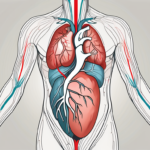
The stethoscope is typically placed on specific areas of the chest, known as auscultatory areas, to listen to different heart sounds. The four primary anatomical areas include the aortic area, pulmonic area, tricuspid area, and mitral area. These regions correspond to the positions of the heart valves and provide access to different sounds produced during cardiac cycles.

The Role of Heart Anatomy in Sound Production

Understanding heart anatomy is crucial for stethoscope placement because it allows healthcare professionals to identify the specific areas where heart sounds can be best heard. For example, the aortic area is located in the second intercostal space on the right sternal border, and is the ideal site to listen to the aortic valve.

The aortic valve is one of the four valves in the heart and is responsible for regulating blood flow from the left ventricle to the aorta, the largest artery in the body. This valve consists of three cusps or leaflets, which open and close with each heartbeat, allowing blood to flow forward and preventing any backward flow.

When listening to the aortic area with a stethoscope, healthcare professionals can detect any abnormalities in the opening and closing of the aortic valve. For example, aortic stenosis, a condition characterized by the narrowing of the aortic valve opening, can produce a distinct heart murmur that can be heard in this area. By carefully placing the stethoscope in the aortic area, healthcare professionals can gather valuable information about the functioning of the aortic valve and the overall health of the patient’s cardiovascular system.

Techniques for Listening to Heart Sounds

Once the basic anatomy and stethoscope placement areas are understood, it is important to use proper techniques for listening to heart sounds. Effective use of the stethoscope is key to accurately detecting and identifying different heart sounds.

Proper Stethoscope Use

When using a stethoscope, it is crucial to ensure a proper seal between the chest and the earpieces. This allows for optimal sound transmission and reduces external noise interference. The diaphragm of the stethoscope is generally used for high-frequency sounds, while the bell is more suited for low-frequency sounds.

Furthermore, it is essential to hold the stethoscope correctly to avoid any unnecessary noise or distortion. The healthcare professional should grasp the earpieces firmly, ensuring a secure fit in the ears. Additionally, the tubing should be positioned comfortably without any kinks or twists that could compromise sound quality.

Identifying Different Heart Sounds

Identifying and differentiating between different heart sounds can be challenging, especially for beginners. With practice and experience, healthcare professionals can learn to recognize normal heart sounds, as well as detect abnormalities such as heart murmurs, clicks, and gallops. Listening to a variety of heart sounds in different clinical scenarios can enhance proficiency in sound interpretation.

One technique that can aid in identifying heart sounds is the use of phonocardiography, which involves recording and analyzing heart sounds using specialized equipment. This advanced technology allows for a more detailed examination of the heart’s acoustic signals, enabling healthcare professionals to detect subtle abnormalities that may not be easily discernible through traditional auscultation alone.

Moreover, it is important to consider the patient’s position during auscultation. Certain heart sounds may be more pronounced or easier to detect when the patient is in a specific position, such as lying on their left side or sitting upright. By adjusting the patient’s position, healthcare professionals can optimize their ability to hear and interpret heart sounds accurately.

Common Challenges in Hearing Heart Sounds

Despite the best placement techniques, there may be challenges in hearing heart sounds clearly. External factors, patient-related factors, and individual factors can all affect the audibility of heart sounds.

Overcoming Obstacles in Heart Sound Detection

To overcome obstacles in heart sound detection, it is important to optimize the listening environment. Minimizing ambient noise, ensuring patient comfort and relaxation, and using proper stethoscope technique can all contribute to clearer heart sound perception.

Tips for Clearer Heart Sound Perception

Here are some tips to improve heart sound perception:

  • Ensure a quiet environment for auscultation
  • Position the patient appropriately for optimal stethoscope access
  • Consider using a higher quality stethoscope for enhanced sound transmission
  • Practice active listening and concentrate on heart sounds to improve recognition

While these tips can greatly improve the ability to hear heart sounds, it is important to acknowledge that there are additional challenges that may arise. One such challenge is the presence of murmurs, which can make it difficult to distinguish between normal heart sounds and abnormal ones.

Murmurs are abnormal sounds that occur during the cardiac cycle and can indicate underlying heart conditions. They can range from being innocent and harmless to being a sign of a serious heart problem. Identifying and interpreting murmurs requires a trained ear and experience in differentiating between various types.

Another challenge in heart sound detection is the presence of artifacts. Artifacts are unwanted sounds or noises that can interfere with the clarity of heart sounds. They can be caused by factors such as patient movement, improper stethoscope placement, or external interference. It is important to be aware of these artifacts and take steps to minimize their impact on auscultation.

Advanced Stethoscope Placement for Specific Conditions

While the basic stethoscope placement techniques are sufficient for most cases, there are instances where specific conditions require advanced placement to enhance sound detection and accuracy.

Adjusting Placement for Heart Murmurs

Heart murmurs are abnormal sounds caused by turbulent blood flow through the heart valves. To better detect heart murmurs, the stethoscope can be positioned in different areas, depending on the suspected valve involved. Adjusting the position can help isolate the specific area where the murmur intensity is the highest.

For example, when suspecting a mitral valve murmur, the healthcare professional may place the stethoscope over the apex of the heart, which is the lower left side of the chest. This position allows for better auscultation of the mitral valve area, where the murmur is most likely to be heard. On the other hand, if an aortic valve murmur is suspected, the stethoscope may be placed at the second intercostal space on the right side of the sternum. This position optimizes the detection of aortic valve abnormalities.

Stethoscope Positioning for Arrhythmias

In cases of arrhythmias, where the heart rhythm is irregular, positioning the stethoscope over the auscultatory areas can help discern the specific pattern of the abnormal heart sounds. By listening in different areas, healthcare professionals can better assess the timing and characteristics of irregular heartbeats.

When evaluating a patient with atrial fibrillation, the healthcare professional may place the stethoscope over the mitral valve area to listen for irregular and rapid heart sounds. This positioning allows for the identification of the characteristic “irregularly irregular” rhythm associated with atrial fibrillation. On the other hand, if ventricular arrhythmias are suspected, the stethoscope may be placed over the left sternal border to focus on the area where abnormal ventricular contractions are more prominent.

By adjusting the stethoscope placement according to the specific condition, healthcare professionals can gather valuable information about the nature and severity of the patient’s cardiovascular abnormalities. This advanced technique enhances diagnostic accuracy and aids in the development of appropriate treatment plans.

Maintaining Your Stethoscope for Optimal Performance

Regular maintenance and care of your stethoscope are essential for preserving its functionality and performance. A well-maintained stethoscope ensures accurate sound transmission and extends the lifespan of your instrument.

Regular Stethoscope Cleaning and Care

To clean your stethoscope, gently wipe it down with an alcohol-based or disinfectant wipe after each use. This helps remove any dirt, oils, or bacteria that may have accumulated on the surface. It is important to avoid using excessive moisture or harsh chemicals, as these can damage the instrument. Instead, opt for a gentle cleaning solution that is specifically designed for stethoscopes.

When cleaning, pay special attention to the ear tips and diaphragm, as these are the areas that come into direct contact with the patient’s skin. Use a soft cloth or cotton swab to clean these parts, ensuring that they are free from any debris or buildup. This not only helps maintain the hygiene of your stethoscope but also ensures optimal sound transmission during auscultation.

Proper Stethoscope Storage

In addition to regular cleaning, proper storage of your stethoscope is crucial for its longevity. After each use, make sure to store your stethoscope in a clean and dry environment. Avoid leaving it exposed to extreme temperatures, direct sunlight, or moisture, as these can lead to deterioration and potential damage to the instrument.

Consider investing in a protective case or pouch to keep your stethoscope safe when not in use. This will help prevent accidental drops, scratches, or any other physical damage that may compromise its performance. By taking these simple storage precautions, you can ensure that your stethoscope remains in optimal condition for years to come.

When to Replace Your Stethoscope

Over time, stethoscopes may undergo wear and tear, reducing their performance. Regularly inspecting your instrument is essential to identify any signs of deterioration. If you notice any cracks, loose parts, or a significant decline in sound quality, it may be time to replace your stethoscope.

Investing in a new stethoscope when needed is crucial for accurate and reliable auscultation. The advancements in stethoscope technology have led to the development of innovative features that enhance sound quality and comfort for both the healthcare professional and the patient. By staying up to date with the latest advancements, you can ensure that you have a stethoscope that meets your specific needs and provides the best possible patient care.

Remember, maintaining your stethoscope is not just about preserving its appearance, but also about ensuring its functionality and accuracy. By following these simple maintenance tips and being proactive in replacing your stethoscope when necessary, you can continue to rely on this essential tool for years to come.

Conclusion

Placing the stethoscope correctly for heart sound detection is fundamental for healthcare professionals. By understanding the anatomy of the heart, using proper techniques, and maintaining your stethoscope, you can enhance your ability to detect and interpret heart sounds accurately. Regular practice and experience are key in developing proficiency in auscultation, allowing for more effective diagnosis and patient care.

what is s3 and s4 heart sounds

Heart sounds are important diagnostic clues that healthcare professionals use to assess the health of a patient’s heart. Two specific heart sounds, known as S3 and S4, play a crucial role in diagnosing various cardiac conditions. Understanding these heart sounds is vital for accurate diagnosis and effective treatment. In this article, we will explore the basics of heart sounds, delve into the characteristics and clinical significance of both S3 and S4 heart sounds, and discuss how to differentiate between them. We will also highlight the challenges in heart sound interpretation and the role of advanced technology in heart sound analysis.

Understanding Heart Sounds

Before diving into the specifics of S3 and S4 heart sounds, let’s first understand the basics of heart sounds. The heart produces distinct sounds during each cardiac cycle, which can be heard using a stethoscope. These sounds are a result of the opening and closing of the heart valves and the blood flow through the chambers.

Heart sounds are typically classified into two components: the first heart sound (S1) and the second heart sound (S2). S1 corresponds to the closure of the mitral and tricuspid valves, while S2 corresponds to the closure of the aortic and pulmonary valves.

The Basics of Heart Sounds

Heart sounds are best heard at specific points on the chest wall, known as auscultatory areas. The S1 sound is heard best at the apex of the heart, while the S2 sound is heard best at the base of the heart.

The S1 heart sound is often described as a “lub” sound, while the S2 heart sound is described as a “dub” sound. These sounds can vary in intensity, pitch, and timing, depending on a person’s age, physical condition, and any underlying cardiac abnormalities.

Additionally, it is important to note that heart sounds can be influenced by external factors such as body position, breathing patterns, and the presence of lung diseases. For example, a person with a respiratory condition may have altered heart sounds due to the increased pressure on the heart caused by difficulty in breathing.

Now, let’s turn our attention to the S3 and S4 heart sounds, which provide additional information about the functioning of the heart.

The Role of Heart Sounds in Diagnostics

Heart sounds serve as valuable diagnostic indicators and can provide insights into a patient’s cardiovascular health. Abnormal heart sounds, such as S3 and S4, can indicate underlying cardiac conditions that warrant further investigation and treatment.

S3 heart sound, also known as the ventricular gallop, is an additional sound that occurs immediately after the S2 heart sound. It is often described as a low-pitched sound resembling the word “Kentucky”. S3 is associated with increased blood flow into the ventricles during the early phase of diastole, indicating decreased ventricular compliance or heart failure.

S4 heart sound, on the other hand, occurs just before the S1 heart sound and is known as the atrial gallop. It is a low-frequency sound that is often described as a “tennessee” or “stiff wall” sound. S4 is associated with increased resistance to ventricular filling, commonly seen in conditions such as hypertension, aortic stenosis, and hypertrophic cardiomyopathy.

By carefully listening to and analyzing heart sounds, healthcare professionals can gather important information about the structure and function of the heart. This, in turn, aids in the diagnosis and management of various cardiovascular conditions.

The S3 Heart Sound

The S3 heart sound, also known as the ventricular gallop, is an extra heart sound that occurs during early diastole. It is best heard with the bell of the stethoscope placed at the apex of the heart.

Characteristics of the S3 Heart Sound

The S3 heart sound is characterized by a low-frequency, dull sound that resembles the word “Kentucky” when auscultated. It occurs immediately after the S2 heart sound and is associated with the rapid filling of the ventricles during early diastole.

When listening to the S3 heart sound, healthcare professionals pay close attention to its timing and intensity. The timing of the S3 heart sound can provide valuable information about the condition of the heart. For example, an early S3 heart sound suggests increased ventricular filling pressure, while a late S3 heart sound may indicate decreased ventricular compliance.

The intensity of the S3 heart sound can also vary, ranging from barely audible to loud and easily detectable. A loud S3 heart sound may suggest a significant increase in ventricular filling pressure, while a soft S3 heart sound may indicate a milder degree of ventricular dysfunction.

The presence of an S3 heart sound may indicate increased blood volume or a weakened left ventricle, commonly seen in conditions such as heart failure or myocardial infarction.

Clinical Significance of the S3 Heart Sound

The S3 heart sound is a clinical sign that healthcare professionals use to assess the severity and progression of heart failure. Its presence can indicate fluid overload or ventricular dysfunction, helping guide treatment decisions and monitor the effectiveness of interventions.

In addition to heart failure, the S3 heart sound can also be present in other conditions, such as dilated cardiomyopathy, valvular regurgitation, and constrictive pericarditis. Therefore, its identification and interpretation are crucial in diagnosing and managing various cardiac disorders.

Patients with an S3 heart sound may require further diagnostic tests, such as echocardiography, to evaluate the underlying cause and determine the best course of action. Echocardiography provides detailed images of the heart’s structure and function, allowing healthcare professionals to assess the extent of ventricular dysfunction, measure ejection fraction, and identify any structural abnormalities.

In summary, the S3 heart sound is an important clinical finding that can provide valuable insights into the functioning of the heart. Its characteristics, timing, and intensity can help healthcare professionals assess the severity of heart failure and guide treatment decisions. Further diagnostic tests may be necessary to determine the underlying cause and develop an appropriate management plan.

The S4 Heart Sound

The S4 heart sound, also known as the atrial gallop, is an additional heart sound that occurs late in diastole. It is best heard with the bell of the stethoscope placed at the apex of the heart.

When listening for the S4 heart sound, healthcare professionals pay close attention to its characteristics. The S4 heart sound is characterized by a low-frequency, stiff, and sharp sound that resembles the word “Tennessee” when auscultated. This unique sound is caused by the contraction of the atria pushing blood into a non-compliant ventricle. It occurs just before the S1 heart sound, which is the normal first heart sound.

The presence of an S4 heart sound may indicate reduced ventricular compliance or increased resistance to ventricular filling. These conditions are commonly seen in patients with hypertension or hypertrophic cardiomyopathy. In hypertension, the heart has to work harder to pump blood against increased resistance in the blood vessels. This increased workload can lead to reduced ventricular compliance, causing the S4 heart sound. Similarly, in hypertrophic cardiomyopathy, the walls of the ventricles become thickened, making it harder for the ventricles to fill with blood. This increased resistance to ventricular filling can also result in the S4 heart sound.

Healthcare professionals use the S4 heart sound as a clinical sign to assess the presence and severity of left ventricular hypertrophy. Left ventricular hypertrophy refers to the thickening of the left ventricle, which can occur as a result of chronic hypertension or other cardiac conditions. By listening for the S4 heart sound, healthcare professionals can gather valuable information about the heart’s structure and function. This information helps guide treatment decisions and monitor disease progression.

If a patient is found to have an S4 heart sound, further diagnostic tests may be required to evaluate the underlying cause. Electrocardiography, or ECG, is commonly performed to assess the electrical activity of the heart and identify any abnormalities. Cardiac imaging, such as echocardiography, may also be used to visualize the heart’s structure and function in more detail. These additional tests help healthcare professionals determine the best course of action for patients with an S4 heart sound.

Differentiating Between S3 and S4 Heart Sounds

Distinguishing between S3 and S4 heart sounds can be challenging, as they occur close to each other in the cardiac cycle. However, there are key differences in sound that can aid in accurate identification.

Key Differences in Sound

One key difference between the S3 and S4 heart sounds is their timing. The S3 heart sound occurs shortly after the second heart sound (S2), while the S4 heart sound occurs just before the first heart sound (S1).

But what causes these differences in timing? Let’s delve deeper into the mechanics of the heart. The S3 heart sound, also known as the ventricular gallop, is caused by the rapid deceleration of blood flow during early diastole. On the other hand, the S4 heart sound, also called the atrial gallop, is caused by the contraction of the atria pushing blood against a stiff ventricle during late diastole.

Another difference between S3 and S4 heart sounds lies in their pitch. The S3 heart sound is typically lower in pitch than the S4 heart sound. This difference in pitch can be attributed to the different mechanisms that produce these sounds. The S3 heart sound is generated by the vibrations of the ventricular walls, while the S4 heart sound is produced by the vibrations of the atrial walls.

Implications for Patient Health

The presence of an S3 or S4 heart sound can provide valuable diagnostic information about a patient’s cardiovascular health. Differentiating between these heart sounds is crucial for accurate diagnosis and appropriate treatment.

When an S3 heart sound is identified, it may indicate underlying heart failure. This could be due to conditions such as dilated cardiomyopathy or volume overload. On the other hand, the identification of an S4 heart sound may suggest the presence of left ventricular hypertrophy. This condition is often associated with chronic hypertension or aortic stenosis.

These findings can guide healthcare professionals in formulating an individualized treatment plan for each patient. For instance, if an S3 heart sound is detected, the focus may be on managing fluid overload and optimizing cardiac function. If an S4 heart sound is present, efforts may be directed towards reducing left ventricular hypertrophy and managing any underlying conditions contributing to its development.

The Importance of Accurate Heart Sound Interpretation

Accurate heart sound interpretation is essential for identifying and managing various cardiac conditions. However, it can pose challenges due to variations in sound characteristics and the presence of additional murmurs or artifacts.

Challenges in Heart Sound Interpretation

One challenge in heart sound interpretation is the ability to differentiate between normal and abnormal sounds. Variations in sound characteristics can make it difficult to distinguish between physiological variations and pathological conditions.

For example, the presence of an S3 heart sound, also known as a ventricular gallop, can be indicative of heart failure or volume overload. This low-frequency sound occurs during early diastole and is caused by rapid filling of the ventricles. On the other hand, an S4 heart sound, also known as an atrial gallop, can suggest decreased ventricular compliance or hypertrophic cardiomyopathy. This high-frequency sound occurs during late diastole and is caused by atrial contraction against a stiff ventricle.

Additionally, the presence of additional murmurs or artifacts, such as lung sounds or extraneous noises, can further complicate the interpretation process. These sounds can mask or mimic abnormal heart sounds, making it challenging to accurately identify and differentiate them.

The Role of Advanced Technology in Heart Sound Analysis

Advanced technology, such as digital stethoscopes and computer-assisted analysis, has revolutionized heart sound analysis. These tools can enhance the accuracy and efficiency of heart sound interpretation, aiding healthcare professionals in making informed diagnostic decisions.

Computer-assisted analysis algorithms can automatically detect and classify abnormal heart sounds, minimizing the risk of human error and providing clinicians with valuable insights. These advancements are particularly beneficial in challenging cases or when expertise is limited.

Furthermore, digital stethoscopes have the ability to amplify and filter heart sounds, allowing for better visualization of subtle abnormalities. They can also record and store heart sounds for further analysis and consultation with specialists, facilitating collaboration and improving patient care.

In conclusion, understanding S3 and S4 heart sounds is vital for healthcare professionals involved in cardiac diagnostics. These unique heart sounds offer valuable information about a patient’s cardiac health and can guide treatment decisions. Accurate interpretation and differentiation between S3 and S4 heart sounds require knowledge, skills, and sometimes the assistance of advanced technology. By utilizing these resources, healthcare professionals can optimize patient care and improve outcomes in cardiac medicine.

how to document heart sounds

Heart sounds are essential diagnostic tools used by healthcare professionals to assess the functioning of the heart. Documenting these sounds accurately is crucial for accurate diagnosis and treatment. In this article, we will explore various aspects of documenting heart sounds, including understanding heart sounds, necessary equipment, listening techniques, accurate documentation, interpreting heart sounds, privacy and ethical considerations, and improving your skills over time.

Understanding Heart Sounds

Before diving into the process of documenting heart sounds, it is vital to have a basic understanding of how these sounds occur. The heart produces two distinct sounds, commonly referred to as S1 and S2.

The Basics of Heart Sounds

Heart sound S1 is the first sound heard during the cardiac cycle and is caused by the closure of the mitral and tricuspid valves. This sound is loudest at the apex of the heart. S2, on the other hand, is the second sound and is caused by the closure of the aortic and pulmonary valves. It is heard most prominently at the base of the heart.

Different Types of Heart Sounds

In addition to S1 and S2, there are other heart sounds that can be indicative of specific cardiac conditions. These additional sounds, such as S3 and S4, can provide valuable information about the functioning of the heart.

S3, also known as the ventricular gallop, is an additional heart sound that can be heard during early diastole. It is caused by the rapid filling of the ventricles and is often associated with conditions such as heart failure. The presence of an S3 sound can indicate a decrease in ventricular compliance and may require further investigation.

S4, on the other hand, is a late diastolic sound that occurs just before S1. It is caused by the contraction of the atria pushing blood into non-compliant ventricles. This sound is often associated with conditions such as hypertension and can indicate decreased ventricular compliance. The presence of an S4 sound can be an important clue in diagnosing certain cardiac abnormalities.

Understanding these additional heart sounds is crucial in accurately assessing and diagnosing cardiac conditions. By listening carefully and identifying the different sounds, healthcare professionals can gather valuable information about the functioning of the heart and provide appropriate treatment.

Necessary Equipment for Documenting Heart Sounds

When it comes to documenting heart sounds, having the right equipment is not just important, it’s vital. Accurate documentation can provide valuable insights into a patient’s cardiovascular health, aiding in diagnosis and treatment. Here, we will delve into two essential tools that every healthcare professional should consider.

Stethoscope Selection

A stethoscope is the quintessential tool for listening to heart sounds, and selecting the right one can make all the difference. When choosing a stethoscope, it is crucial to prioritize sound quality and comfort. Opt for a high-quality stethoscope that is specifically designed to capture the subtle nuances of heart murmurs, allowing you to detect even the faintest abnormalities. Additionally, ensure that the stethoscope fits comfortably in your ears, as a poor fit can lead to distorted sound transmission and compromise your ability to accurately document heart sounds.

Recording Devices for Heart Sounds

While a stethoscope is undoubtedly the primary tool for auscultation, considering the use of recording devices can greatly enhance your ability to document heart sounds. These devices can serve as invaluable aids, allowing for the playback and analysis of recorded heart sounds. By capturing the audio, you can meticulously review the recordings, focusing on specific segments or comparing them to previous auscultations. This not only facilitates accurate documentation but also enables you to track any changes in the heart sounds over time, providing a comprehensive picture of the patient’s cardiovascular health.

Moreover, recording devices can be particularly beneficial in educational settings. They allow instructors to share real-life examples of heart sounds with students, fostering a deeper understanding of cardiac auscultation. Students can listen to the recordings repeatedly, honing their skills and becoming more proficient in identifying various heart murmurs and abnormalities.

When it comes to documenting heart sounds, precision and attention to detail are paramount. By carefully selecting a high-quality stethoscope and considering the use of recording devices, healthcare professionals can ensure accurate and comprehensive documentation, ultimately leading to improved patient care.

Techniques for Listening to Heart Sounds

Now that you have the necessary equipment ready, it’s time to explore the techniques for effectively listening to heart sounds. Listening to the heart can provide valuable information about a person’s cardiovascular health, and mastering the art of auscultation is an essential skill for healthcare professionals.

One important aspect of listening to heart sounds is proper stethoscope placement. Placing the stethoscope’s diaphragm over the appropriate areas of the chest is crucial to capture the heart sounds accurately. The diaphragm should be in direct contact with the skin and positioned firmly but gently. This ensures that the sound waves produced by the heart are transmitted clearly to the stethoscope, allowing for a more accurate assessment of the heart’s function.

However, it’s not just about where you place the stethoscope; it’s also about how you interpret the sounds you hear. Developing the ability to distinguish normal and abnormal heart sounds is essential. Normal heart sounds are regular and consistent, reflecting the normal functioning of the heart’s valves and chambers. On the other hand, abnormal sounds may indicate underlying cardiac conditions, such as murmurs, valve disorders, or even heart failure.

Identifying abnormal heart sounds requires a keen ear and a deep understanding of the different types of murmurs and their characteristics. Continuous learning and practice are crucial for honing this skill. Healthcare professionals often spend countless hours listening to heart sounds, attending workshops, and studying various resources to improve their diagnostic abilities.

By mastering the techniques for listening to heart sounds, healthcare professionals can gather valuable information about a patient’s cardiovascular health. This information can aid in the diagnosis and management of various cardiac conditions, ultimately improving patient outcomes. So, take the time to perfect your stethoscope placement, train your ears to differentiate between normal and abnormal sounds, and never stop learning in this fascinating field of cardiology.

Documenting Heart Sounds Accurately

To ensure accurate documentation of heart sounds, healthcare professionals should follow specific guidelines.

Describing Heart Sounds

When documenting heart sounds, it is essential to provide a concise and detailed description. Include information such as timing, intensity, pitch, quality, and any other relevant characteristics.

For example, when describing the timing of heart sounds, note whether they occur during systole or diastole. This information can help identify potential abnormalities in the cardiac cycle. Additionally, it is crucial to document the intensity of heart sounds, whether they are faint or loud, as this can provide valuable insights into the overall cardiac function.

Importance of Consistent Terminology

Using consistent terminology when documenting heart sounds helps ensure clear and effective communication among healthcare professionals. Familiarize yourself with standard terminology and utilize it consistently in your documentation.

One commonly used term in describing heart sounds is “murmur.” A murmur refers to an abnormal sound heard during the cardiac cycle, which can indicate underlying heart conditions. It is important to specify the characteristics of a murmur, such as its location, timing, and intensity, to assist in accurate diagnosis and treatment planning.

Furthermore, consistency in terminology extends beyond individual healthcare professionals. It is crucial for medical institutions to establish standardized terminology guidelines to promote uniformity in documentation. This allows for easier comparison of patient records and facilitates research and data analysis.

Interpreting Heart Sounds

Interpreting heart sounds is a skill that requires knowledge and experience. Here are two critical aspects to consider:

Recognizing Common Heart Conditions through Sounds

Through careful listening and interpretation, certain heart conditions can be identified by their distinct sound patterns. Familiarize yourself with these patterns to aid in diagnosis and treatment decisions.

One common heart condition that can be recognized through sound is mitral valve regurgitation. This condition occurs when the mitral valve does not close properly, allowing blood to flow backward into the left atrium. When listening to the heart sounds, you may hear a distinct “whooshing” sound during systole, indicating the regurgitation of blood.

Another condition that can be identified through heart sounds is aortic stenosis. This occurs when the aortic valve becomes narrowed, obstructing the flow of blood from the left ventricle to the aorta. As a result, you may hear a harsh, systolic ejection murmur during auscultation, indicating the presence of aortic stenosis.

When to Refer for Further Evaluation

While healthcare professionals can gather valuable information from heart sounds, it is essential to recognize when to refer patients for further evaluation by specialists. An accurate diagnosis often requires a comprehensive assessment and additional diagnostic tests.

One instance where referral for further evaluation is necessary is when a patient presents with a heart murmur that is accompanied by symptoms such as chest pain, shortness of breath, or fainting. These symptoms may indicate a more serious underlying heart condition that requires specialized care.

Additionally, if the heart sounds are unclear or if there is uncertainty in the interpretation, it is crucial to refer the patient to a cardiologist. Cardiologists have the expertise and specialized equipment to perform advanced diagnostic tests, such as echocardiography, to obtain a more detailed assessment of the heart’s structure and function.

Privacy and Ethical Considerations

When documenting heart sounds, healthcare professionals must adhere to privacy and ethical guidelines.

Ensuring patient consent and confidentiality is of utmost importance when recording heart sounds for documentation purposes. Before capturing any audio, it is crucial to obtain explicit consent from the patient. This not only respects their autonomy but also establishes a foundation of trust between the healthcare provider and the patient. By obtaining consent, patients are actively involved in their own care, empowering them to make informed decisions about their medical information.

Protecting patient confidentiality goes beyond obtaining consent. It involves implementing robust security measures to safeguard the privacy of sensitive medical records. Healthcare professionals must ensure that all records, including heart sound recordings, are stored securely and accessible only to authorized individuals. This means utilizing encrypted storage systems, employing strict access controls, and regularly auditing the handling of patient data.

Patient Consent and Confidentiality

Obtain patient consent before recording heart sounds for documentation purposes. Protect patient confidentiality by ensuring that all records are stored securely and accessible only to authorized individuals.

Respecting patient privacy extends beyond the realm of legal obligations. It is an ethical imperative that healthcare professionals must uphold. By maintaining strict confidentiality, healthcare providers demonstrate their commitment to the well-being and dignity of their patients.

Ethical Guidelines for Heart Sound Documentation

Follow ethical guidelines established by professional bodies when documenting heart sounds. These guidelines aim to protect the rights and well-being of patients and ensure professionalism in the practice.

Professional bodies, such as medical associations and regulatory authorities, have developed ethical guidelines specifically tailored to heart sound documentation. These guidelines serve as a compass, guiding healthcare professionals in their practice and ensuring that they adhere to the highest standards of ethical conduct.

Some of the key ethical considerations outlined in these guidelines include respecting patient autonomy, maintaining confidentiality, and avoiding any form of exploitation or harm. Healthcare professionals are expected to approach heart sound documentation with empathy, sensitivity, and a commitment to the well-being of their patients.

Improving Your Skills Over Time

Documenting heart sounds accurately is a skill that can be honed with continuous learning and practice. The journey to becoming proficient in this field requires dedication and a commitment to staying updated with the latest research and techniques in cardiac auscultation.

Continuous Learning and Practice

One of the key aspects of improving your skills in documenting heart sounds is to stay updated with the latest advancements in the field. Attending workshops, conferences, and webinars can provide valuable opportunities to enhance your knowledge and skills. These events bring together experts in the field who share their insights and experiences, allowing you to learn from the best.

Moreover, continuous learning also involves staying informed about the latest research findings in cardiology. As new studies emerge, they shed light on different aspects of heart sounds and provide valuable insights into diagnosing and treating various cardiac conditions. By keeping up with the latest research, you can incorporate new knowledge and techniques into your practice, ensuring that you are providing the best possible care to your patients.

Staying Updated with Latest Research and Techniques

Advancements in auscultation technology are constantly being made, and it is crucial to stay informed about these developments. New tools and devices are being introduced to improve the accuracy and efficiency of heart sound documentation. By familiarizing yourself with these advancements, you can ensure that you are utilizing the most effective equipment in your practice.

Additionally, staying updated with the latest research findings allows you to gain a deeper understanding of heart sounds and their significance in diagnosing cardiac conditions. New studies may reveal previously unknown correlations between specific heart sounds and certain diseases, enabling you to make more accurate diagnoses and provide targeted treatment plans.

By understanding heart sounds, acquiring the necessary equipment, utilizing proper listening techniques, documenting accurately, interpreting sounds, considering privacy and ethical aspects, and continuously improving your skills, you can become proficient in documenting heart sounds. This expertise will contribute to more accurate diagnoses and improved patient outcomes.

where to auscultate heart sounds

Auscultating, or listening to the heart sounds, is a fundamental skill for healthcare professionals. It provides vital information about the condition of the heart and helps in diagnosing various cardiac abnormalities. To effectively auscultate heart sounds, it’s crucial to know where to listen and understand the significance of each auscultation point. In this article, we will explore the key aspects of heart auscultation, including understanding heart sounds, tools for auscultation, the anatomy of the heart and auscultation points, techniques for auscultation, interpreting heart sounds, common challenges, and tips for improving auscultation skills.

Understanding Heart Auscultation

The Basics of Heart Sounds

Heart sounds are the noises generated by the functioning of the human heart. They are produced by the closing of heart valves during the cardiac cycle. The primary sounds heard during auscultation are the first and second heart sounds (S1 and S2). S1 corresponds to the closure of the mitral and tricuspid valves, while S2 coincides with the closure of the aortic and pulmonary valves.

Heart murmurs, which are additional sounds during the cardiac cycle, may indicate abnormalities such as valve malfunctions or structural defects. Identifying and understanding these sounds is crucial in making accurate diagnoses.

Importance of Accurate Heart Auscultation

Accurate heart auscultation plays a vital role in diagnosing and monitoring heart conditions. It helps in assessing heart valve functions, detecting abnormal heart rhythms, and identifying other cardiac abnormalities. By listening to the heart sounds at specific auscultation points, healthcare professionals can gather valuable information about the patient’s cardiac health and make informed decisions regarding treatment.

When performing heart auscultation, healthcare professionals use a stethoscope to listen to the sounds produced by the heart. The stethoscope consists of a chest piece, which is placed on the patient’s chest, and earpieces, which allow the healthcare professional to hear the sounds clearly. The chest piece contains a diaphragm and a bell, each designed to capture different types of sounds.

During auscultation, the healthcare professional carefully moves the stethoscope across different areas of the chest to listen to specific heart sounds. The sounds produced by the heart can vary depending on the position of the patient, the presence of underlying conditions, and other factors. Therefore, it is essential for the healthcare professional to have a thorough understanding of the normal and abnormal heart sounds to accurately interpret what they hear.

In addition to auscultation, other diagnostic tests such as echocardiograms and electrocardiograms may be used to further evaluate the heart’s structure and function. These tests provide detailed images and measurements that can aid in the diagnosis and treatment of various cardiac conditions.

Tools for Heart Auscultation

Stethoscope Selection and Use

The stethoscope is the primary tool used for heart auscultation. Selecting the appropriate stethoscope is essential for obtaining clear and accurate sounds. Different stethoscope models offer varying levels of sound amplification and ambient noise reduction, so it’s important to choose one suited to your needs. Additionally, using the stethoscope correctly, positioning it on the patient’s chest, and adjusting the earpieces properly are crucial for optimal sound transmission.

Advanced Auscultation Devices

Besides traditional stethoscopes, advanced auscultation devices, such as electronic stethoscopes and digital auscultation systems, are gaining popularity. These devices offer enhanced sound amplification, noise cancellation, and the ability to record auscultation findings for further analysis. While traditional stethoscopes remain the gold standard, these advanced devices can be helpful in certain situations, such as in noisy environments or when additional features are necessary.

Electronic stethoscopes, for example, are designed with built-in amplifiers that can significantly enhance the sound quality. This is particularly beneficial when listening to faint heart murmurs or abnormal heart sounds that may be difficult to detect with a traditional stethoscope. The amplification feature allows healthcare professionals to better assess and diagnose cardiac conditions, leading to more accurate treatment plans.

Digital auscultation systems, on the other hand, take auscultation to a whole new level by incorporating advanced technology. These systems consist of a digital stethoscope connected to a computer or mobile device, which allows for real-time visualization of heart sounds. The visual representation of the heart sounds can aid in identifying subtle abnormalities or patterns that may not be easily discernible by listening alone. Furthermore, digital auscultation systems often come with software that enables the recording and storage of auscultation findings, facilitating collaboration and continuity of care.

Anatomy of the Heart and Auscultation Points

The Four Heart Valves

The human heart is a remarkable organ that plays a vital role in circulating oxygen-rich blood throughout the body. It consists of four valves, each with its own unique position and function in regulating the flow of blood. These valves are like gatekeepers, allowing blood to flow in one direction while preventing any backward flow.

The first valve we will explore is the mitral valve, located between the left atrium and the left ventricle. It is composed of two flaps, or cusps, that open and close synchronously to ensure the proper flow of blood from the atrium to the ventricle.

The tricuspid valve, on the other hand, is situated between the right atrium and the right ventricle. As its name suggests, it consists of three cusps that work together to prevent the backflow of blood from the ventricle to the atrium.

Moving on to the aortic valve, it is positioned at the exit of the left ventricle, where it connects to the aorta, the largest artery in the body. This valve has three cusps that open and close with each heartbeat, allowing the oxygenated blood to be pumped out of the heart and into the systemic circulation.

Lastly, we have the pulmonary valve, which is located at the exit of the right ventricle. It has three cusps that open and close to facilitate the flow of deoxygenated blood from the heart to the lungs, where it can be replenished with oxygen.

Identifying Auscultation Points

Now that we have a better understanding of the four heart valves, let’s delve into the importance of identifying the auscultation points. Auscultation, which involves listening to the sounds produced by the heart using a stethoscope, is a crucial diagnostic tool in assessing the heart’s health and detecting any abnormalities.

The auscultation points, also known as cardiac landmarks, are specific locations on the chest where the sounds of the heart are best heard. These points serve as guideposts for healthcare professionals to accurately assess the functioning of the heart valves and detect any potential issues.

The aortic area, located in the second intercostal space to the right of the sternum, is one of the key auscultation points. By placing the stethoscope in this area, healthcare providers can listen for the characteristic sounds associated with the opening and closing of the aortic valve.

The pulmonic area, situated in the second intercostal space to the left of the sternum, is another important auscultation point. Here, healthcare professionals can listen for the sounds produced by the pulmonary valve, which provide valuable insights into the functioning of the right side of the heart.

The tricuspid area, found in the lower left sternal border, is where the sounds of the tricuspid valve can be best heard. By carefully auscultating this area, healthcare providers can assess the integrity of the valve and identify any abnormalities that may affect blood flow.

Lastly, the mitral area, located in the fifth intercostal space at the midclavicular line, allows healthcare professionals to listen for the sounds produced by the mitral valve. This valve is of particular interest as it is responsible for preventing the backflow of blood from the left ventricle to the left atrium.

Accurate identification of these auscultation points and the use of appropriate techniques are essential in ensuring a precise assessment of the heart sounds. By carefully listening to the heart’s symphony, healthcare providers can gain valuable insights into the functioning of the heart valves and provide the best possible care for their patients.

Techniques for Auscultating Heart Sounds

Patient Positioning for Optimal Auscultation

The positioning of the patient during heart auscultation can significantly impact the quality of sounds heard. Positioning the patient correctly, such as in a seated or supine position, ensures the heart is appropriately aligned for optimal listening. This alignment allows healthcare professionals to accurately assess the heart sounds and detect any abnormalities that may be present.

However, patient positioning is not limited to a simple seated or supine position. In some cases, certain maneuvers can be employed to further enhance the clarity of heart sounds. For instance, asking the patient to take deep breaths can help accentuate specific heart sounds, especially those associated with valvular abnormalities. This technique allows healthcare professionals to gather more detailed information about the patient’s cardiac health.

Systematic Auscultation Approach

Adopting a systematic approach to auscultation ensures that all relevant areas are carefully assessed. Following a specific sequence, such as starting from the aortic area and moving clockwise around the chest, helps ensure consistency and accuracy in assessing various heart sounds. This systematic approach not only aids in the identification of abnormalities but also allows for the comparison of sound characteristics at different auscultation points.

During the systematic auscultation process, healthcare professionals pay close attention to the different areas of the heart, including the aortic, pulmonic, tricuspid, and mitral areas. By meticulously evaluating each of these areas, they can identify any deviations from the normal heart sounds and determine the underlying causes. This comprehensive assessment is crucial in providing accurate diagnoses and developing appropriate treatment plans for patients.

Interpreting Heart Sounds

Understanding heart sounds is a fundamental skill for healthcare professionals, especially when it comes to differentiating between normal and abnormal sounds. While normal heart sounds can vary slightly from person to person, they generally exhibit specific characteristics that can be identified through careful auscultation. These characteristics include intensity, duration, and pitch.

Normal heart sounds, often referred to as “lub-dub,” are produced by the closing of the heart valves. The first sound, known as S1, is caused by the closure of the mitral and tricuspid valves, while the second sound, S2, is produced by the closing of the aortic and pulmonary valves. These sounds are typically heard as a rhythmic, synchronized pattern, indicating the proper functioning of the heart.

Normal Heart Sounds

Familiarizing yourself with the typical features of normal heart sounds enables you to identify any deviations from the norm during auscultation. For example, the intensity of the heart sounds can vary depending on factors such as age, body size, and overall cardiovascular health. In general, a louder S1 sound may indicate a forceful closure of the mitral and tricuspid valves, while a louder S2 sound may suggest increased pressure within the aorta and pulmonary artery.

The duration of normal heart sounds is also important to consider. Normally, S1 is longer and more pronounced than S2, with a brief pause between the two sounds. However, in certain conditions, such as aortic stenosis, the duration of S2 may be prolonged, indicating a narrowing of the aortic valve.

Pitch, another characteristic of heart sounds, can provide valuable information about the condition of the heart. A higher-pitched S1 sound may indicate a stiffening of the mitral and tricuspid valves, while a higher-pitched S2 sound may suggest increased blood flow velocity within the aorta and pulmonary artery.

Abnormal Heart Sounds and What They Mean

While normal heart sounds follow a predictable pattern, abnormal heart sounds can indicate various cardiac abnormalities. These abnormal sounds, such as murmurs or additional heart sounds, can provide valuable insights into the underlying condition.

Murmurs, for example, are abnormal sounds caused by turbulent blood flow within the heart. They can be indicative of conditions such as valvular diseases, ventricular septal defects, or aortic regurgitation. The timing, loudness, pitch, and quality of the murmur can help healthcare professionals diagnose and monitor these specific cardiac disorders.

Additional heart sounds, such as S3 and S4, can also be abnormal findings. S3, commonly heard in conditions like congestive heart failure, is caused by rapid ventricular filling during the early phase of diastole. S4, on the other hand, is often associated with conditions like hypertrophic cardiomyopathy and is caused by the contraction of the atria against a stiff ventricle.

Interpreting these abnormal heart sounds requires a keen ear and a thorough understanding of cardiac physiology. By carefully analyzing the timing, loudness, pitch, and quality of these sounds, healthcare professionals can make accurate diagnoses and provide appropriate treatment for patients with cardiac abnormalities.

Common Challenges in Heart Auscultation

Overcoming Background Noise

Auscultation, the process of listening to the sounds produced by the heart using a stethoscope, can be quite challenging, especially in noisy clinical environments. The presence of background noise can interfere with the accuracy of auscultation, making it difficult for healthcare professionals to identify and interpret the subtle sounds of the heart.

To overcome background noise, healthcare professionals employ various techniques. One effective approach is to ask the patient to sit in a quiet area, away from distractions and external disturbances. This creates an environment conducive to accurate auscultation, allowing the healthcare professional to focus on the sounds of the heart without any interference.

In addition to the patient’s positioning, the correct placement of the stethoscope is crucial. Healthcare professionals are trained to position the stethoscope correctly on the patient’s chest, ensuring optimal sound transmission and minimizing the impact of external noise. This precise placement enables them to capture the heart sounds with greater clarity and accuracy.

Furthermore, advancements in technology have led to the development of auscultation devices with noise-reducing features. These devices are designed to filter out unwanted background noise, enhancing the quality of auscultation. By utilizing such devices, healthcare professionals can further minimize the impact of background noise, ensuring that the heart sounds are heard clearly and accurately.

Dealing with Patient Discomfort

While background noise poses a challenge in heart auscultation, patient discomfort or anxiety can also affect the accuracy of the procedure. It is not uncommon for patients to feel uneasy or anxious during a medical examination, especially when it involves the heart, a vital organ that holds great significance.

Healthcare professionals play a crucial role in addressing patient discomfort and ensuring a positive patient experience during heart auscultation. Creating a calming environment is essential, as it helps to alleviate the patient’s anxiety. Simple measures such as dimming the lights, playing soothing music, or providing a comfortable seating arrangement can contribute to a more relaxed atmosphere.

Moreover, effective communication between the healthcare professional and the patient is vital. Explaining the procedure in a clear and concise manner helps to demystify the process, easing the patient’s concerns. By providing information about what to expect, healthcare professionals can help patients feel more at ease and reduce any unnecessary anxiety.

Establishing a good rapport with the patient is equally important. Demonstrating empathy, actively listening to their concerns, and addressing any questions they may have can go a long way in building trust and creating a positive patient-provider relationship. Maintaining professionalism throughout the auscultation process ensures that the patient feels respected and valued, further enhancing their overall experience.

In conclusion, heart auscultation presents its own set of challenges, including background noise and patient discomfort. By employing techniques to overcome background noise and creating a calming environment for patients, healthcare professionals can enhance the accuracy of auscultation and provide a positive patient experience.

Tips for Improving Auscultation Skills

Continuous Learning and Practice

Auscultation is a skill that improves with practice and experience. Healthcare professionals should dedicate time for continuous learning and honing their auscultation skills. Regular practice with normal and abnormal heart sounds helps develop a keen ear for detecting subtle variations and enhances diagnostic accuracy.

Utilizing Auscultation Simulations and Recordings

Advancements in technology have enabled the creation of auscultation simulations and recordings. These tools allow healthcare professionals to practice their auscultation skills in a controlled environment while providing realistic heart sound representations. Utilizing such simulations and recordings can be an effective way to improve proficiency in differentiating normal and abnormal heart sounds.

Mastering the art of heart auscultation is essential for accurate diagnosis and monitoring of cardiac conditions. By understanding heart sounds, utilizing the appropriate tools, knowing the anatomy and auscultation points, applying proper techniques, and continuous practice, healthcare professionals can enhance their auscultation skills and provide optimal care to their patients.

where to listen to heart sounds

Listening to heart sounds is an essential skill for healthcare professionals, particularly those in cardiology. Heart sounds can provide valuable insights into a person’s cardiovascular health and help identify potential issues. In this article, we will explore the different areas of the chest where heart sounds can be heard and the significance of listening to them.

Understanding Heart Sounds

Before we dive into the specifics of where to listen to heart sounds, let’s first understand the basics of heart sounds. The normal heartbeat produces two distinguishable sounds, commonly referred to as S1 and S2.

S1 is the first heart sound, also known as “lub.” It is caused by the closure of the mitral and tricuspid valves, marking the beginning of systole, the contraction phase of the heart. S2 is the second heart sound, known as “dub.” It occurs when the aortic and pulmonic valves close at the end of systole, signifying the start of diastole, the relaxation phase of the heart.

The Basics of Heart Sounds

S1 and S2 are the primary heart sounds heard during a normal heartbeat. Understanding these sounds and their frequency is crucial in recognizing abnormal heart sounds, which may indicate underlying cardiovascular conditions.

However, it is important to note that heart sounds can vary depending on factors such as age, body size, and physical activity. For example, in children, the heart sounds may be louder and higher pitched compared to adults. Additionally, athletes may have slower heart rates, resulting in a more pronounced S2 sound.

Furthermore, by familiarizing themselves with normal heart sounds, healthcare professionals can more effectively interpret and diagnose abnormalities that may arise during auscultation, or the process of listening to heart sounds. This skill is particularly valuable in emergency situations, where quick and accurate assessment of heart sounds can help determine the appropriate course of action.

The Importance of Listening to Heart Sounds

Listening to heart sounds can provide valuable information about a person’s cardiovascular health. Heart murmurs, extra sounds, or irregular rhythms can be indicative of various conditions, such as valve disorders, heart failure, or congenital heart defects.

Moreover, regular auscultation of heart sounds allows healthcare professionals to monitor changes in a patient’s condition over time. By comparing current and previous heart sounds, they can identify any concerning developments and adjust treatment plans accordingly.

It is worth mentioning that advancements in technology have also enhanced the accuracy and efficiency of heart sound analysis. Digital stethoscopes and computerized algorithms can now assist healthcare professionals in detecting subtle abnormalities that may have been missed in the past.

In conclusion, understanding heart sounds and their significance is essential in the field of cardiology. By honing their auscultation skills and staying updated with the latest diagnostic tools, healthcare professionals can provide optimal care for patients with cardiovascular conditions.

Anatomy of the Heart and Its Sounds

Understanding the anatomical locations where specific heart sounds can be best heard is key in proper auscultation techniques. Let’s explore the four main areas of the chest where heart sounds are primarily heard:

The Four Main Areas to Listen to Heart Sounds

  1. The Aortic Area: This area, located in the second intercostal space at the right sternal border, is the best location to hear the aortic valve closure, corresponding to the second heart sound (S2).
  2. The Pulmonary Area: Situated in the second intercostal space at the left sternal border, this area is optimal for detecting the pulmonary valve closure, also known as the second heart sound (S2).
  3. The Tricuspid Area: Found in the fourth intercostal space at the left sternal border, this area allows for auscultation of the tricuspid valve closure, corresponding to the first heart sound (S1).
  4. The Mitral Area: Located at the fifth intercostal space at the midclavicular line, this area is best for detecting the mitral valve closure, representing the first heart sound (S1).

Identifying Normal Heart Sounds

Recognizing normal heart sounds is crucial in distinguishing abnormal ones. Establishing a baseline of what is considered normal for each individual patient is vital for accurate diagnosis and assessment.

Normal heart sounds should be clear and well-defined, with S1 and S2 occurring in a regular rhythm. Additionally, the intensity, pitch, and duration of each sound play a role in their identification.

Let’s delve deeper into the fascinating world of heart sounds. Each heart sound is a result of a complex interplay between the valves, chambers, and blood flow within the heart. Understanding the intricate mechanisms behind these sounds can provide valuable insights into the overall health of the cardiovascular system.

When the aortic valve closes, it produces the second heart sound (S2) in the aortic area. This closure prevents blood from flowing back into the left ventricle, ensuring efficient circulation throughout the body. The pulmonary valve, on the other hand, closes to prevent blood from flowing back into the right ventricle, creating the second heart sound (S2) in the pulmonary area.

In the tricuspid area, the tricuspid valve closes to prevent blood from flowing back into the right atrium. This closure generates the first heart sound (S1), marking the beginning of ventricular contraction. Similarly, in the mitral area, the mitral valve closes to prevent blood from flowing back into the left atrium, producing the first heart sound (S1).

When listening to heart sounds, it is essential to pay attention to the timing and characteristics of each sound. The intensity of the sounds can vary depending on factors such as the thickness of the chest wall and the position of the patient. The pitch of the sounds can range from high-pitched to low-pitched, providing valuable clues about the underlying conditions of the heart.

By carefully analyzing the duration of each heart sound, healthcare professionals can gather crucial information about the efficiency of the heart’s pumping action. Abnormalities in the duration of heart sounds can indicate conditions such as valve stenosis or regurgitation, which require further investigation and intervention.

Remember, auscultation is an art that requires practice and a keen ear. By familiarizing yourself with the anatomy of the heart and its sounds, you can enhance your ability to detect abnormalities and provide optimal care for your patients.

Tools for Listening to Heart Sounds

To listen to heart sounds accurately, healthcare professionals utilize specific tools. The most common instrument for auscultation is the stethoscope, but digital devices are also becoming increasingly popular.

Using a Stethoscope

The stethoscope is a primary tool for listening to heart sounds. It consists of a chest piece, which contains a diaphragm and a bell, and two earpieces connected by tubing. When placed on the patient’s chest, the diaphragm and bell transmit the sound vibrations to the ears of the healthcare professional.

Using a stethoscope requires skill and precision. Healthcare professionals must position the chest piece correctly to capture the heart sounds effectively. They need to apply the right amount of pressure to the chest piece, ensuring a clear transmission of sound. Additionally, they must listen attentively, focusing on different areas of the chest to detect any abnormalities or murmurs.

By mastering the technique of using a stethoscope, healthcare professionals can enhance their ability to detect and analyze heart sounds accurately. They can pick up subtle nuances in the sounds, such as murmurs, clicks, or extra heart sounds, which can provide valuable diagnostic information.

Digital Devices for Heart Sound Monitoring

Advancements in technology have led to the development of digital devices capable of recording and analyzing heart sounds. These devices often utilize sensors and algorithms to provide detailed information about the condition of the heart.

With digital devices, healthcare professionals can not only listen to heart sounds but also visualize them in real-time. Some devices even offer the ability to record and store the sounds for further analysis or comparison. This can be particularly useful for long-term monitoring or telemedicine purposes, allowing healthcare professionals to assess heart sounds remotely.

While digital devices can be beneficial, it is important for healthcare professionals to ensure that they are reliable and produce accurate results. Regular calibration and maintenance of these devices are necessary to ensure their precision. Additionally, healthcare professionals must be knowledgeable about the limitations and potential pitfalls of digital auscultation, as it may not always replace the skill and experience required for accurate diagnosis.

Techniques for Listening to Heart Sounds

Listening to heart sounds requires more than just having the right tools. Proper techniques and positioning are also crucial in capturing the most accurate and useful information.

Positioning for Optimal Listening

To optimize auscultation, it is essential to have the patient in the appropriate position. The patient should be sitting upright, as this allows gravity to assist in the movement of blood and enhances sound transmission.

Furthermore, the healthcare professional should ensure a quiet environment to minimize external noise interference, which could affect the clarity of the heart sounds. This can be achieved by closing doors and windows, turning off any unnecessary equipment, and politely asking individuals in the vicinity to maintain silence during the examination.

Another aspect to consider is the patient’s comfort. Ensuring that they are relaxed and at ease can help reduce any potential tension or anxiety that may affect their heart rate or rhythm, ultimately impacting the accuracy of the auscultation.

Differentiating Heart Sounds

Distinguishing between different heart sounds can be challenging, especially for those new to auscultation. Developing a discerning ear requires regular practice and exposure to a variety of patients with differing heart conditions.

One helpful technique is to listen for the S1 and S2 sounds, which are the primary heart sounds. The S1 sound is produced by the closure of the mitral and tricuspid valves, while the S2 sound is created by the closure of the aortic and pulmonary valves. By familiarizing oneself with the timing and characteristics of these sounds, healthcare professionals can better identify abnormalities.

Additionally, familiarizing oneself with the characteristics of abnormal heart sounds, such as murmurs or gallops, is vital in identifying potential issues and referring patients for further evaluation when necessary. These abnormal sounds can provide valuable clues about underlying heart conditions, such as valve disorders or heart muscle abnormalities.

Furthermore, it is important to consider the patient’s medical history and any associated symptoms when interpreting heart sounds. Understanding the context in which the sounds are heard can aid in making a more accurate diagnosis and determining the appropriate course of action.

Interpreting Heart Sounds

Interpreting heart sounds is a skill that requires both knowledge and experience. It involves recognizing any abnormalities or deviations from normal heart sounds, which may have clinical implications.

When listening to heart sounds, healthcare professionals pay close attention to the timing, intensity, and quality of the sounds. The normal heart has two distinct sounds, often referred to as “lub” and “dub.” The first sound, “lub,” is caused by the closure of the mitral and tricuspid valves, while the second sound, “dub,” is caused by the closure of the aortic and pulmonary valves. These sounds are usually crisp and clear, with a regular rhythm.

Recognizing Abnormal Heart Sounds

Abnormal heart sounds, such as extra heart sounds or murmurs, can be indicative of underlying cardiac conditions. These sounds may suggest problems with heart valves, the presence of a defect, or an inefficient pumping mechanism.

For example, a common abnormal heart sound is a heart murmur. Murmurs are often described as a whooshing or swishing sound that occurs between the normal lub-dub sounds. They can be caused by turbulent blood flow through narrowed or leaking heart valves. Healthcare professionals use their expertise to differentiate between innocent murmurs, which are harmless, and pathological murmurs, which may require further investigation and treatment.

By accurately identifying abnormal heart sounds, healthcare professionals can guide further diagnostic investigations and develop appropriate treatment plans. This may include ordering additional tests such as echocardiograms, electrocardiograms, or stress tests to assess the structure and function of the heart.

When to Seek Medical Advice

Although healthcare professionals are trained to listen to heart sounds, it is crucial for individuals to be vigilant about their own cardiovascular health. If you experience any concerning symptoms like chest pain, shortness of breath, or palpitations, it is imperative to seek medical advice promptly.

Furthermore, it is important to note that not all heart conditions manifest with obvious symptoms. Some individuals may have underlying heart problems without experiencing any noticeable signs. Regular check-ups and screenings can help detect these hidden conditions and prevent potential complications.

Early detection and appropriate management can significantly contribute to improved outcomes and overall heart health. Remember, your heart is a vital organ that deserves attention and care. By staying informed and proactive, you can take control of your cardiovascular well-being and lead a heart-healthy life.

Learning Resources for Heart Sounds

For those interested in further expanding their knowledge and expertise in auscultation, numerous learning resources are available that can enhance their understanding of heart sounds.

Online Platforms for Learning Heart Sounds

The internet offers various online platforms dedicated to teaching heart sounds. These platforms often provide interactive tutorials, audio recordings, and case studies that allow healthcare professionals to practice and refine their auscultation skills.

One popular online platform is “HeartSoundQuiz,” which offers a comprehensive collection of heart sound recordings accompanied by detailed explanations. This interactive tool allows users to listen to different heart sounds and test their knowledge by identifying the specific sounds they hear. With its user-friendly interface and extensive library of cases, “HeartSoundQuiz” provides a valuable resource for healthcare professionals seeking to improve their auscultation skills.

Books and Guides on Heart Sounds

Traditional resources, such as books and guides, continue to be valuable tools for learning about heart sounds. These resources often provide in-depth explanations, diagrams, and real-life examples that aid in understanding the nuances of heart sounds.

“The Art of Auscultation: Mastering Heart Sounds” by Dr. Emily Thompson is a highly recommended book for healthcare professionals looking to deepen their understanding of heart sounds. This comprehensive guide covers a wide range of topics, including the anatomy of the heart, the physiology of heart sounds, and the interpretation of abnormal sounds. With its clear and concise explanations, accompanied by detailed illustrations, this book serves as an invaluable resource for both beginners and experienced practitioners.

In addition to books, pocket guides are also popular among healthcare professionals for quick reference. “Auscultation Made Easy: A Pocket Guide to Heart Sounds” by Dr. Michael Johnson is a compact yet comprehensive guide that provides a systematic approach to understanding and recognizing heart sounds. With its concise explanations and mnemonic techniques, this pocket guide is a practical resource that can be easily carried and consulted during clinical practice.

Continual education and practice are necessary to refine these skills and stay up to date with advancements in the field. Whether through online platforms or traditional resources, healthcare professionals can enhance their aptitude in auscultation, ultimately benefiting their patients and improving their own clinical practice.